- Therapeutic Cataract & Refractive
- Lens Technology
- Glasses
- Ptosis
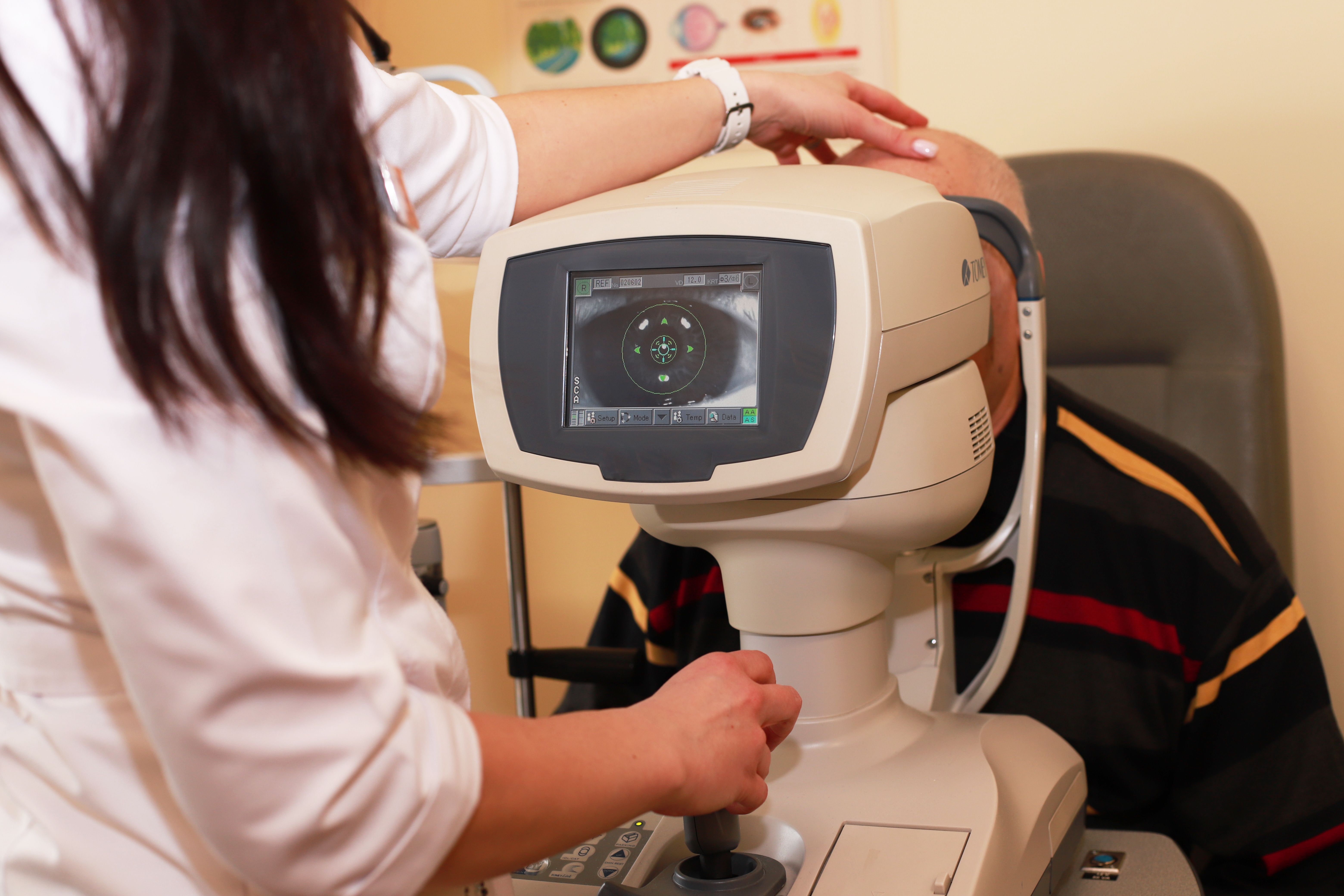
- Comprehensive Eye Exams
- AMD
- COVID-19
- DME
- Ocular Surface Disease
- Optic Relief
- Geographic Atrophy
- Cornea
- Conjunctivitis
- LASIK
- Myopia
- Presbyopia
- Allergy
- Nutrition
- Pediatrics
- Retina
- Cataract
- Contact Lenses
- Lid and Lash
- Dry Eye
- Glaucoma
- Refractive Surgery
- Comanagement
- Blepharitis
- OCT
- Patient Care
- Diabetic Eye Disease
- Technology
Identifying and treating glaucoma in high myopes
Risk factors take priority


Millions of individuals worldwide have glaucoma, with 80 million having been estimated to be affected by the disease.
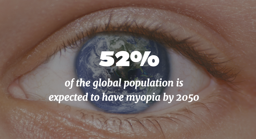
Another disease that is running rampant is myopia. Since the beginning of the 21st century, the incidence rates have been increasing alarmingly by leaps and bounds—approximately by 6% a decade.
Research shows that the incidence increased from 22% in 2000 to 33% in 2020.1 By 2050, an incidence rate of 52% is projected.
Unfortunately, glaucoma and myopia go hand in hand. Myopia is a risk factor for the development of glaucoma. A meta-analysis of 11 population-based studies showed that patients with myopia have a doubling of the risk of developing glaucoma.2
The caveat here is that the 2 diseases can be difficult to differentiate, Ruth Williams, MD, pointed out at the 26th Annual Glaucoma Symposium. She is from the Wheaton Eye Clinic, Chicago, and on the Board of Directors of the Glaucoma Research Foundation.
High myopes can present a confounding picture, in that they can have intraocular pressures in the normal range but abnormal imaging results and retinal nerve fiber layer thicknesses that are outside the normal range.
Optic nerve morphology and visual fields
In both diseases, the optic nerves can resemble each other. Patients with high myopia have characteristic axial elongation and tilting of the nerve, which can look like glaucoma. The picture can be confusing if the intraocular pressure falls within the normal range.
Glaucoma is characterized by a large cup, asymmetry, nerve fiber layer loss, vertical cup elongation, notching of the nerve, disc hemorrhage, peripapillary atrophy, and nasalization of vessels.
Similarly, myopia is characterized by a large disc, increased vertical diameter, temporal tilt, shallow cupping, increased beta zone peripapillary atrophy, and decreased retinal nerve fiber layer thickness.


The visual field defects in myopia can include an enlarged blind spot, a nasal step in 15.6%, and arcuate and paracentral scotomas in 35.5% and 28.1%, respectively. Arcuate scotomas, Dr. Williams noted, may correspond to thin rim tissue at the inferior and superior poles.
So optical coherence imaging may not be very helpful.
Important treatment questions
So the question arises for physicians about when to begin treatment when faced with a patient suspected of having glaucoma and concomitant myopia.
She advises physicians to assess each patient’s risk factors. These include the family history, the presence of higher intraocular pressure, a thin cornea, and low blood pressure. The next step in the assessment is identifying progression in these patients.
However, when considering treatment she advised avoiding filtering surgery.
A second question that arises concerns myopia treatment as a glaucoma prevention strategy. Treatment may be a consideration, since the data from a recent study indicated that for every 1-diopter increase in myopia, the prevalence of open-angle glaucoma increases by 20%.3
When faced with differentiating glaucoma and myopia, Dr. Williams reviewed the primary considerations.
- 1. Structural and functional defects in myopic eyes are difficult to distinguish from glaucomatous defects.
- 2. Visual defects and OCT imaging may not be helpful for differentiating the 2 diseases.
- 3. Patients should be monitored for progression.
- 4. Patient risk factor analysis is especially important.
She concluded, “Physicians should monitor patients for progression and lean toward treatment in the presence of risk factors, such as age, family history, thin cornea, higher intraocular pressure, and low blood pressure.”
References
1. Holden BA, Fricke TR, Wilson DA, et al. Global prevalence of myopia and high myopia and temporal trends from 2000 through 2050. Ophthalmology 2016;123:1036-42.
2. Marcus MW, de Vries MM, Junoy Montolio FG, et al. Myopia as a risk factor for open-angle glaucoma: A systemic review and meta-analysis. Ophthalmology 2011;118:1989-94.e2.
3. Bullimore MA, Ritchey ER, Shah S, et al. The risks and benefits of myopia control. Ophthalmology 2021;128:1561-79.