
Differential diagnoses: How to determine cause of visual field loss
Management decisions for a patient who presents with a visual field defect should never be made before considering a host of other etiologies that can masquerade as glaucoma by the type of visual field loss.
New York-Management decisions for a patient who presents with a visual field defect (VFD) should never be made before considering a host of other etiologies that can masquerade as glaucoma by the type of visual field loss (VFL), said Sherry J. Bass, OD, FAAO, at International Vision Expo East.


Consider big picture


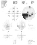
VFL associated with glaucoma presents initially as paracentral defects with progression to nasal step defects, arcuate and Bjerrum defects, altitudinal defects, and ultimately, if the disease is not controlled, peripheral field constriction. For each of these types of defects, there are different etiologies to consider as an alternative diagnosis.
Non-glaucomatous etiologies of paracentral field loss include hereditary macular disease, such as Stargardt macular degeneration and cone dystrophy, and optic pits. Superior paracentral VFLs can be found in patients with a hereditary macular disease as a result of their typical use of superior eccentric fixation to achieve better vision.
In patients with an optic pit, usually located at the inferior temporal border of the optic nerve head, paracentral field loss is secondary to loss of the retinal nerve fiber layer between the macula and optic nerve that can exist in this condition, Dr. Bass explained.
Nasal field defects are also a sign of early glaucoma but can alternatively be caused by optic disc drusen. Diagnosis of optic disc drusen may be challenged by the fact that the lesions may be buried in younger patients.
"The finding of an optic nerve head with blurry disc borders and no cupping in a patient with a nasal field defect should suggest the diagnosis of optic nerve drusen," Dr. Bass said.
"Further evaluation should include B-scan ultrasonography," she continued. "However, this imaging is only effective for identifying drusen that are calcified, which includes about 50% of disc drusen."
Newsletter
Want more insights like this? Subscribe to Optometry Times and get clinical pearls and practice tips delivered straight to your inbox.


 of patients believe that medical technology innovations will enhance care for the aging population, with 90% of providers recognizing the need to address specific requirements for older patients. Image credit: AdobeStock/buritora")

