How OCT can help a smaller practice
Spectral domain optical coherence tomography (SD-OCT) has grown in importance to optometric patient care. One OD shares her experience of moving from quarterly use of a rented device to purchasing an OCT for her small private practice and how it has improved clinical care and patient education.

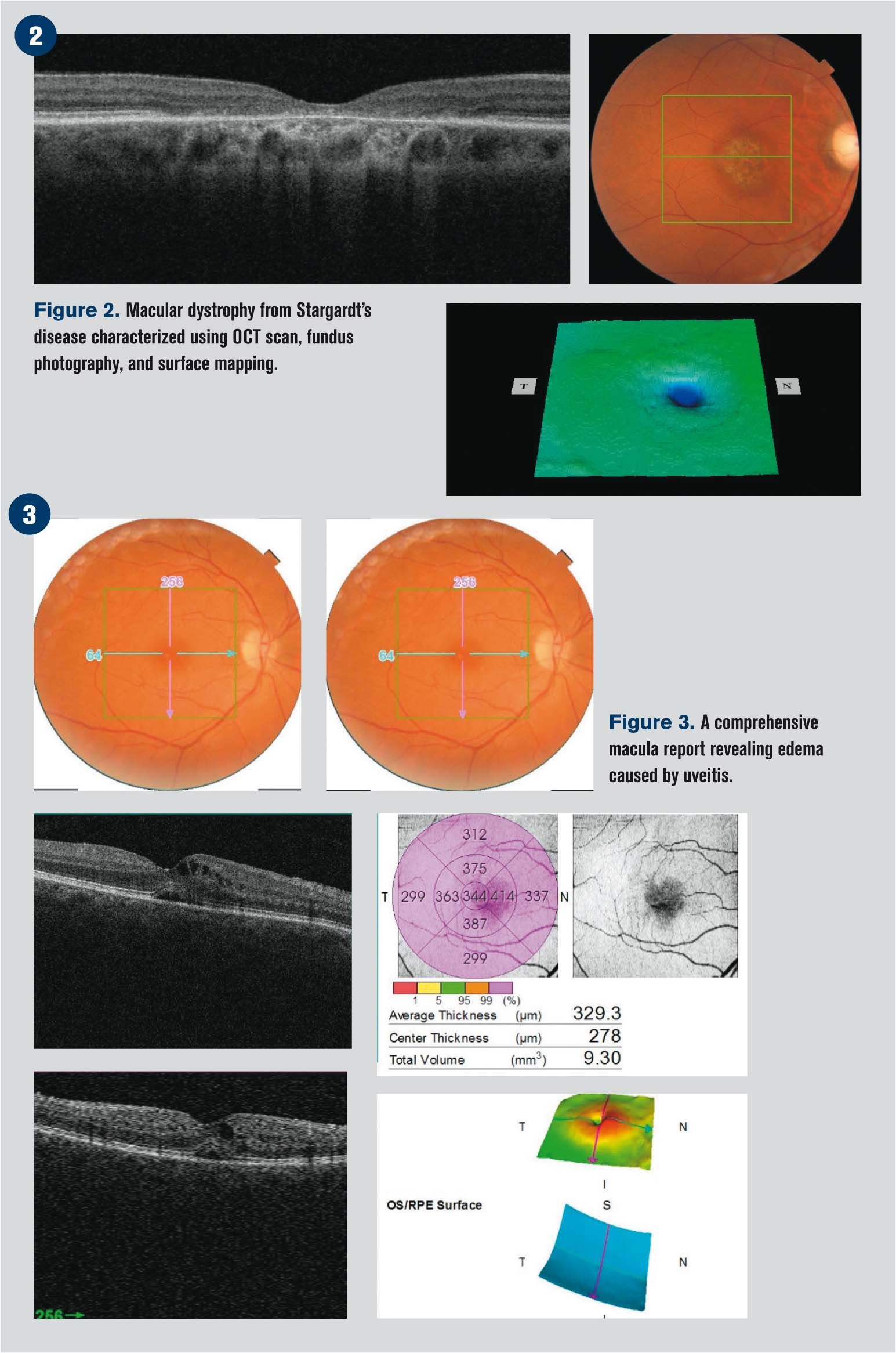
Spectral domain optical coherence tomography (SD-OCT) is rapidly becoming a must-have device for optometrists. OCT is the standard of care for detecting diabetic macular edema and is the fastest, least invasive method for detecting fluid in the retina.1-4 It has also become a game-changer for monitoring macular degeneration, glaucoma, and diabetic retinopathy.5-7
ODs in smaller practices may have the challenge of justifying the expense of this technology.
My practice situation
In my 28-year optometric career, I have worked in multiple settings from big-box retailers to ophthalmology clinics to private optometry practices. When I decided to take what I had learned and open cold to start my own solo practice, one of the more intimidating aspects of the process was acquiring the required technology.
I knew would need an OCT, but initially I was not able to afford one. For the first few years, I rented a mobile OCT service on a quarterly basis which came to my practice for two to three hours with a technician.
Related: Affording OCT in your practice
As I watched my waiting room become increasingly full with longer waiting times on OCT days, I realized that this method was not providing the best patient experience. Continuity of care was often lost because I had to ask patients to return on another day for an OCT to further investigate a finding. Additionally, urgent scans had to be sent out and done elsewhere. I was quickly recognizing that I had to have my own OCT.
Practice-changing addition
In shopping for an instrument, the office space required was a big consideration. Two devices made the short list, Topcon 3D SD-OCT-1 Maestro and Zeiss Cirrus HD-OCT. Due to its smaller footprint, the Maestro was literally a better fit.

In our small office, the Maestro and monitor sit on a two-instrument table which is conveniently pushed up against a wall. The instrument’s feature of a pivoting viewing screen pivot allows the patient and technician to be on the same side of the instrument table.
When I bought my OCT, I found that it immediately changed my patient care. Before my in-office OCT, I would describe to the patient what I saw, why I was asking him to return for follow up when the OCT was in the office, or why I was referring him elsewhere.
Related: OCT in pediatric eye disease
Now, if I see something of concern, I say, “It looks like you have <insert condition here>. We’re going to take a scan of that today, and I’ll show you.” I walk the patient to the OCT to immediately obtain the imaging I need.
Having the device in my practice rather than renting a mobile device has enhanced my patients’ confidence in me. Both new and established patients have commented on how they are impressed by technology to monitor their conditions.
Educating patients about their eyes gives them knowledge to better understand their condition. I like to tell patients that I need their help in caring for their eyes-I show them their ocular condition, not simply point to a picture in a book.

Such personal imagery is especially important for glaucoma patients-they need to know why I’m asking them to use eye drops every day.
For retina patients, I want them to understand exactly why I am sending them to a specialist and what it means for their vision when they have fluid in the macula.
Related: 6 OCT pitfalls to avoid
Using OCT in my practice also enables me to make sure my referrals are appropriate, and I have found that specialists appreciate that efficiency. They are confident in my reports to them because I have pre-scanned my patients, and I can make timely referrals knowing a patient has a disease which needs advanced attention. Other specialists, such as rheumatologists, send me referrals for baseline studies in patients who take long-term medications and for urgent care when sudden change of vision occurs in patients experiencing autoimmune disease flare-ups.
Power of knowledge
When my patients return for their follow-up OCTs, they know exactly what I’m looking for, and they are more engaged in their care. This is facilitated partly because of the device’s design-it fits into a small space, and the screen is reversible. At my office, I am side-by-side with the patient. When the image pops up on the screen, we look at it together, and I take her through what we are seeing. I can show her the optic nerve, blood vessels, and point out the macula.
In a macular degeneration patient, for example, I would point out early drusen and explain drusen it is why I have recommended he take supplements and use an Amsler grid at home. This gives me an opportunity to review heart-healthy lifestyle recommendations and the importance of UV protection.
When patients see those little yellow dots and know they mean early macular degeneration, they instantly understand more about their condition. I didn’t realize the impact on patients of seeing their eyes on the OCT screen. Patients ask wonderful questions and show a stronger interest in their eye care. If I find and point out an urgent condition, they get it; they understand why they need follow-up appointments.
Long-term care
A sizable percentage of our patients are business travelers, relocating every two to four years. Many others are snowbirds. I remind patients that their OCT scans and photos are part of their permanent health record-if they move, their next doctor can pick up where I left off in following their ocular health. Patients appreciate this.
Our EHR has an online patient portal, which is helpful for such traveling patients. Having access to their records 24/7 has been ideal for patient convenience and long-term continued health care.
Wrapping up
Newer, small-footprint SD-OCT technology can be a win-win for patients and doctors. I’m excited to see where the next generation of OCT technology takes practitioners. I encourage clinicians with small practices to consider taking the plunge and investing by purchasing or leasing their own OCT system to have a detailed view of what’s going on inside their patients’ eyes.
Related: Identifying common macular conditions with OCT
References
1. Kang SW, Park CY, Ham DI. The correlation between fluorescein angiographic and optical coherence tomographic features in clinically significant diabetic macular edema. Am J Ophthalmol. 2004 Feb;137(2):313-22.
2. Kim BY, Smith SD, Kaiser PK. Optical coherence tomographic patterns of diabetic macular edema. Am J Ophthalmol. 2006 Sep;142(3):405-412.
3. Otani T, Kishi S, Maruyama Y. Patterns of diabetic macular edema with optical coherence tomography. Am J Ophthalmol. 1999 Jun;127(6):688-693.
4. Panozzo G, Parolini B, Gusson E, Mercanti A, Pinackatt S, Bertoldo G, Pignatto S. Diabetic macular edema: an OCT-based classification. Semin Ophthalmol. 2004 Mar-Jun;19(1-2):13-20.
5. Chen TC. Spectral domain optical coherence tomography in glaucoma: qualitative and quantitative analysis of the optic nerve head and retinal nerve fiber layer (an AOS thesis). Trans Am Ophthalmol Soc. 2009 Dec;107:254-281.
6. Kagemann L, Wollstein G, Ishikawa H, Bilonick RA, Brennen PM, Folio LS, Gabriele ML, Schuman JS. Identification and assessment of Schlemm’s canal by spectral domain optical coherence tomography. Invest Ophthalmol Vis Sci. 2010 Aug;51(8):4054-4059.
7. Pekmezci M, Porco TC, Lin SC. Anterior segment optical coherence tomography as a screening tool for the assessment of the anterior segment angle. Ophthalmic Surg Lasers Imaging. 2009 Jul-Aug;40(4):389-398.
Newsletter
Want more insights like this? Subscribe to Optometry Times and get clinical pearls and practice tips delivered straight to your inbox.