The importance of multidisciplinary care for diabetes
Recently, a colleague wrote me to express his concern about a primary care physician (PCP) in his community acquiring digital retinal photographs of his diabetes patients. One of those patients presented to the optometrist’s office with the impression that “all he needed was a refraction” since the PCP had “already checked him for diabetic retinopathy.”

Recently, a colleague wrote me to express his concern about a primary care physician (PCP) in his community acquiring digital retinal photographs of his diabetes patients. One of those patients presented to the optometrist’s office with the impression that “all he needed was a refraction” since the PCP had “already checked him for diabetic retinopathy.”
Related: What we can learn from managing diabetic patients
This made me wonder how many other optometrists are facing this predicament. I suspect this is a circumstance eyecare providers may increasingly encounter as economic pressures push insurers and providers to efficiently deliver care and consolidate control over the breadth and scope of health care encounters (i.e. do more with less money and fewer visits).
Dilated ophthalmoscopy instead of a screening
Part of the argument for “diabetic retinopathy screening” derives from the fact that significant minorities of people with diabetes still are not receiving recommended annual dilated eye examinations by optometrists and ophthalmologists.1
In addition, there also may be the perception that rates of severe diabetic retinopathy are low, an argument countered by studies showing that as many as 1 in 20 adults with diabetes have vision-threatening retinopathy2 (nearly 1 in 15 Latino Americans, and 1 in 10 African Americans).

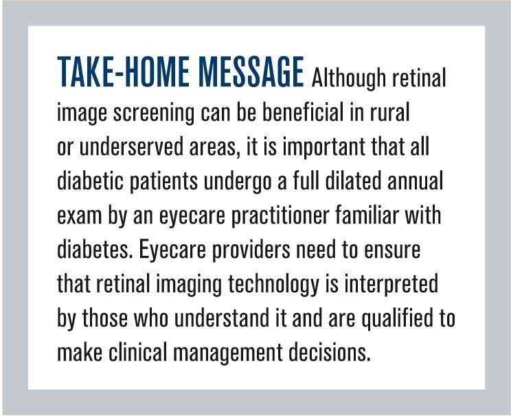
I totally advocate digital retinal imaging for purposes of identifying diabetic retinopathy in underserved populations, provided these images are interpreted by trained observers.
Moreover, this technology is invaluable for patient education, documentation of findings, and sometimes even staging and diagnosis of disease. (Let’s face it, some patients are more tolerant than others when it comes to letting us scrutinize their retinas during dilated fundus examination.
Finding any/every microaneurysm in a photophobic patient with diabetic retinopathy can be challenging, and red-free digital imaging can be enormously helpful even to experienced clinicians).
However, we all know that conventional digital retinal imaging is no substitute for dilated ophthalmoscopy in patients with diabetes for a number of very good reasons.
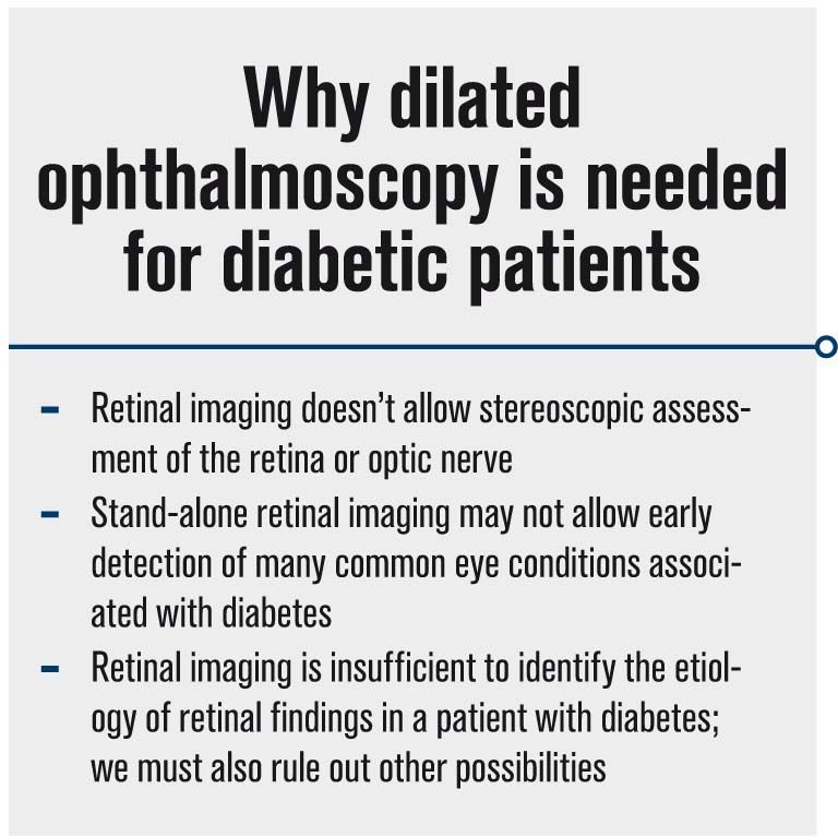
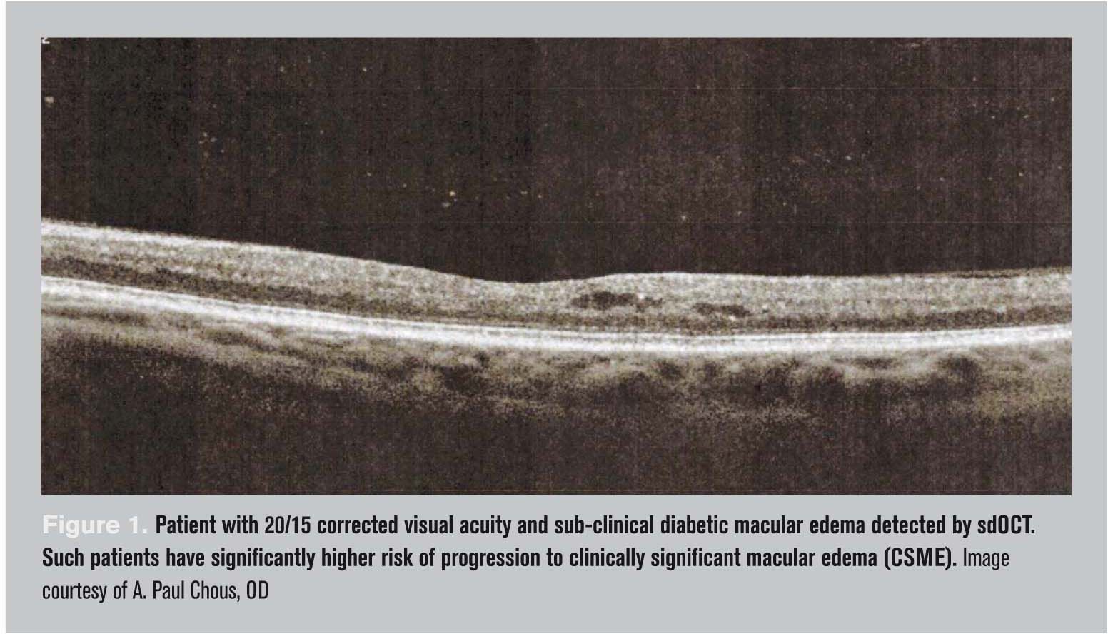
First, retinal imaging doesn’t allow stereoscopic assessment of the retina or optic nerve. We frequently see patients with diabetic macular edema or glaucoma but exhibiting relatively normal-appearing fundus images. In the absence of stereoscopic exam and/or OCT, these cases would not be diagnosed or managed appropriately.3
Second, as a stand-alone technique, retinal imaging may not allow early detection of many common eye conditions associated with diabetes, including cataract, glaucoma, ocular surface disease, lid disease, refractive fluctuation signaling unstable diabetes control, and cranial nerve paresis and palsy. Every optometrist and ophthalmologist knows there is much more to diabetic eye disease than “just” diabetic retinopathy.4
Related: Diabetic eye diseases projected to increase
Third, retinal imaging is insufficient to identify the etiology of retinal findings in a patient with diabetes; we must also rule out other possibilities (e.g. hypertension, venous occlusive disease, even ocular ischemic syndrome) with trained and practiced clinical acumen.
Instruments and their associated technologies do not make diagnoses, nor do they exercise clinical judgment-only an experienced and knowledgeable eyecare specialist, skilled with both the acquisition and the interpretation of the information gleaned from those instruments and technologies, can make the right diagnosis and use sound clinical judgment based on the entirety of patient information, including case history.
Next: Detecting DR in under-served areas

There are some very good public health reasons to employ digital retinal imaging systems for the detection of diabetic retinopathy, particularly sight-threatening diabetic retinopathy (proliferative disease and diabetic macular edema) and especially in under-served areas in the U.S. and internationally.
In fact, the Joslin Vision Network has demonstrated the effectiveness of population-based diabetic retinopathy screening using digital photography,5,6 but it must be remembered that these images are interpreted by highly trained readers who interface with eyecare specialists available to confirm disease severity and examine and treat patients.
However, even these valuable programs have their limitations: They are not designed to detect ocular diabetes complications aside from retinopathy or other overt retinal or optic nerve disease. They are not designed to prevent diabetes-related eye disease but rather to detect vision-threatening retinopathy and prevent catastrophic vision loss.
They are not geared to deliver the kind of patient education available from a face-to-face encounter with an eye doctor, especially with respect to emerging preventative strategies for ocular and systemic diabetes complications as well as new and emerging forms of treatment.
Knowledge is power, and patients with diabetes need plenty of both. There are many excellent reasons, above and beyond the detection of vision-threatening retinopathy, to get an annual dilated eye examination by a diabetes-savvy optometrist.
Next: Multidisciplinary care benefits patients

Multidisciplinary care benefits patients
We frequently see patients who have both diagnosed and undiagnosed diabetes as well as ocular complications of diabetes. Sometimes we are the only healthcare professionals these patients have seen in years, which puts us in the position to refer to and collaborate with primary care providers and other specialists who manage diabetes and its complications.
An eye examination represents an opportunity for us to educate patients about preventing diabetes, the meaning and importance of glycosylated hemoglobin (HbA1c), good blood pressure and blood lipid control, and even measure these in our offices (though I believe it is imperative to let patients know the vital importance of confirmation and follow-up with their PCPs or endocrinologists).
This not only represents an opportunity to save patients’ vision but, sometimes, their lives. Moreover, there is demonstrable benefit when diabetes patients hear mutually reinforcing messages about good self-care from their various healthcare providers, including their eye doctors.7,8

What might I suggest to our colleague who encountered the patient who came to believe that he needed nothing more than a refraction, that he did not require a dilated eye examination from the eye doctor after having his retinas photographed by the (presumably well-intentioned) PCP?
My recommendation is to proactively educate both this patient and every patient we see about why digital retinal photography is no substitute for thorough eye examination by an eyecare specialist, any more than a dentist listening to your heart with a stethoscope before extracting a tooth is substitute for thorough cardiac evaluation by a cardiologist.
This is especially true in at-risk patients-and every patient with diabetes is at-risk.
Related: Diabetes nation-addressing an epidemic
Next: Recommendations for the family physician
Recommendations for the family physician
As for the family physician, I would recommend calling and writing him to make the following points:
• Two-dimensional images of the retina may not allow detection of diabetic macular edema, the leading cause of vision loss in patients with type 2 diabetes;9 optometrists have the tools and experience (OCT, stereoscopic fundus lenses used through a dilated pupil) that allow us to make this critical determination; Figure 1).
• Diabetic retinopathy often occurs outside the field of view afforded by most fundus cameras;10 optometrists have the tools and experience to detect this (dilated fundus exam, ultra-wide field imaging; Figure 2).
• Diabetes affects the eyes in a myriad of ways, including increased risk of glaucoma and other optic neuropathies, cataract, corneal disease, subtle cranial neuropathy, refractive error instability reflecting sub-optimal glycemic control; eye doctors are trained and experienced to diagnose and manage these problems.
Drug Topics: Best apps for managing diabetes
• Tell the PCP you appreciate her commitment to excellent patient care, but fundus photography is no substitute for a comprehensive dilated eye exam by a knowledgeable and experienced eye care provider, and you, the primary eyecare specialist, want to help the PCP deliver the recognized standard of care to her patients.

This should represent a great opportunity for optometrists to build collaborative relationships with primary care, internal medicine, and endocrinology. Being direct and confident will oftentimes lead to productive conversations and improved relationships with other healthcare providers built upon mutual respect and trust.
There will likely be increasing numbers of primary care physicians and other non-ophthalmic providers using digital retinal imaging. This is potentially a good thing that may identify retinal catastrophes and facilitate immediate and necessary eye care.
We must, however, strongly advocate for our patients and our profession to ensure that this technology is not misused or misunderstood by the general public or providers who are not qualified to make or, as importantly, to dismiss important diagnoses and/or exercise proper clinical decision-making based on training and experience.
Click here to check out the latest news and advice on diabetes
References
References:
1. Sloan FA, Yashkin AP, Chen Y. Gaps in receipt of regular eye examinations among medicare beneficiaries diagnosed with diabetes or chronic eye diseases.
Ophthalmology. 2014 Dec;121(12):2452-60.
2. Zhang X, Saaddine JB, Chou CF, et al. Prevalence of diabetic retinopathy in the United States, 2005-2008. JAMA. 2010 Aug 11;304(6):649-56.
3. Hirano T, Iesato Y, Toriyama Y, Imai A, Murata T. Detection of fovea-threatening diabetic macular edema by optical coherence tomography to maintain good vision by prophylactic treatment. Ophthalmic Res. 2014;52(2):65-73.
4. Jeganathan VS1, Wang JJ, Wong TY. Ocular associations of diabetes other than diabetic retinopathy. Diabetes Care. 2008 Sep;31(9):1905-12.
5. Cavallerano AA, Cavallerano JD, Katalinic P, et al. A telemedicine program for diabetic retinopathy in a Veterans Affairs Medical Center--the Joslin Vision Network Eye Health Care Model. Am J Ophthalmol. 2005 Apr;139(4):597-604.
6. Cavallerano JD, Silva PS, Tolson AM, et al. Imager evaluation of diabetic retinopathy at the time of imaging in a telemedicine program. Diabetes Care. 2012 Mar;35(3):482-4.
7. Wagner H, Pizzimenti JJ, Daniel K et al. Eye on diabetes: a multidisciplinary patient education intervention. Diabetes Educ. 2008 Jan-Feb;34(1):84-9.
8. Sundling V, Gulbrandsen P, Jervell J, Straand J. Care of vision and ocular health in diabetic members of a national diabetes organization: a cross-sectional study. BMC Health Serv Res. 2008 Jul 28;8:159.
9. Klein R, Klein BEK, Moss SE, Cruickshanks KJ. The Wisconsin epidemiologic study of diabetic retinopathy. XV: the long-term incidence of macular edema. Ophthalmology 1995;102:7–16.
10. Silva PS, Cavallerano JD, Sun JK, et al. Peripheral lesions identified by mydriatic ultrawide field imaging: distribution and potential impact on diabetic retinopathy severity. Ophthalmology. 2013 Dec;120(12):2587-95.
Newsletter
Want more insights like this? Subscribe to Optometry Times and get clinical pearls and practice tips delivered straight to your inbox.