Treating blepharitis in the pediatric population
When your pediatric patient presents with irritated, itchy eyelids with reddened lid margins, diagnosing blepharitis may be the easy part of patient care. Implementing a treatment regimen for patients who are infants, toddlers, or school-age children, requires optometrists to use not only their knowledge but their clinical art of practice as well.

When your pediatric patient presents with irritated, itchy eyelids with reddened lid margins, diagnosing blepharitis may be the easy part of patient care. Implementing a treatment regimen for patients who are infants, toddlers, or school-age children, requires optometrists to use not only their knowledge but their clinical art of practice as well.
The telltale signs of blepharitis are not likely to be missed.
Common ocular findings include bilateral eyelid inflammation, vessel telangiectasia, and hard, fibrinous, crusts and scales with occasional misdirected or missing lashes.1
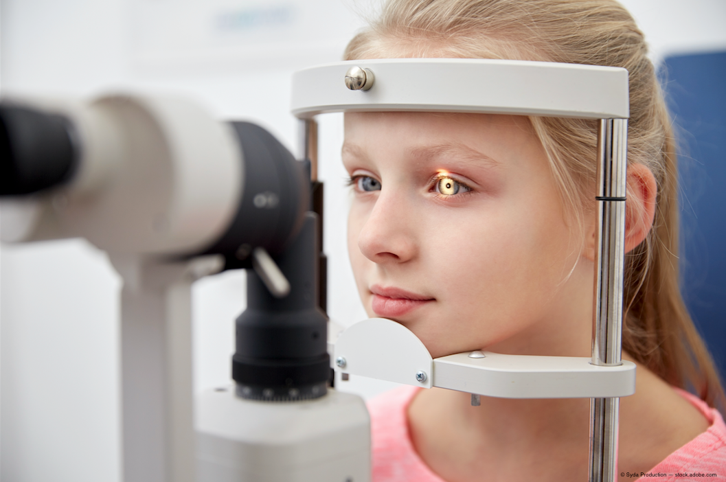
Anterior segment examination-with a slit lamp and/or transilluminator and magnifying lens-is important because many patients (even those old enough to talk) will not report symptoms. Often diagnosis is made based on findings alone.2,3
Related: Pros and cons of available MGD treatments
The onset of blepharitis usually occurs between the ages of six to 10 years. The prevalence of pediatric blepharitis is thought to be on the rise, due to the increase in pediatric contact lenses wear, particularly orthokeratology lenses, in this population.4
Based upon the anatomical location of the inflammation, there are two broad types of blepharitis: anterior and posterior.
Anterior blepharitis involves the skin of the “outer lid” and lashes, while posterior blepharitis involves the cutaneous mucous junction of the lid and as well as meibomian glands.5,6 Both types commonly occur in children, with anterior blepharitis-characterized by flakes and debris along the skin and lashes-being slightly more frequent than posterior blepharitis, that often manifests as meibomian gland dysfunction (MGD).7
Related: In blepharitis, expert looks to restore eye's natural balance
Causes
The most frequent causes of blepharitis include:
• Overgrowth of bacteria, most commonly Staphylococcus species
• Seborrheic dermatitis characterized by dandruff of the scalp, eyebrows, and skin
• Clogged or malfunctioning meibomian glands
• Combination of two or more of these etiologies
MGD occurs as meibomian gland secretions thicken and become clogged due to inflammation, obstruction, and/or gland atrophy. The clinical picture of blepharitis is thought to be the result of several factors, including eyelid gland secretions, bacterial flora, and in some cases, immune dysfunction in the form of Type 3 hypersensitivity reactions to bacterial exotoxins.1,8,9
Less common causes of blepharitis include:
• Rosacea, a skin condition characterized by facial redness
• Mite infestation of the eyelash follicles and meibomian glands
• Herpes simplex infection
Course of treatment
Clinically, with good patient compliance, most cases of pediatric blepharitis resolve in one to two weeks, when the initial findings are mild to moderate.
Differences may exist between different pediatric populations. It has been reported in Asia that pediatric patients have a more severe presentation and that clinical resolution requires a longer period of management.10
In cases of recalcitrant blepharitis, or when the patient presents with atypical findings that do not fit the profile of blepharitis, clinicians should reconsider their working diagnosis and determine if a less common cause is the culprit.11
Fortunately, a good history, observation, and anterior segment evaluation will often be enough to make a diagnosis of blepharitis. Tests and procedures used in ocular surface disease management in adults, such as Schirmer tests, tear osmolarity, and meibomian gland expression are not likely to be successful when the patient is a child.
Whenever possible, use of sodium fluorescein (and possibly other vital dyes) should be attempted to assist in evaluating potential lid changes and meibomian gland and tear quality and corneal integrity. Based on the overall severity of clinical findings, an assessment of severity can be made and management adjusted accordingly
Most patients will respond to a tier approach: lid hygiene measures should be initiated first, with topical medications added next, and when necessary, prescribing systemic medications
Related: Incorporating meibomian gland imaging
Eyelid hygiene
Lid hygiene is the core treatment in blepharitis. It is important to stress that blepharitis is a chronic condition and that treatment must be maintained even after symptoms improve to avoid a relapse.
Many times the directions for lid hygiene will incorporate a three-step approach with heat and massage targeting meibomian gland complications, lid cleaning targeting debris removal from the eyelid skin and lashes, and artificial tears following the other management for generalized removal of debris from the cul-de-sac. Each step can be enhanced or modified based on the specific findings of a patient and the severity of disease.
Heat is particularly important for posterior blepharitis because it softens meibomian gland secretions and breaks up dried discharge. Lid cleaning is essential for anterior blepharitis because it reduces the overall bacterial population and the associated exotoxins. Due to the fact that clinically, blepharitis may have contributions from multiple causes, a lid hygiene routine usually incorporates both heat and cleansing.12
Milder cases of blepharitis may resolve with lid hygiene alone. A good start is to apply a clean washcloth soaked in warm water on top of closed eyelids for five to 10 minutes, one to two times daily. Implementing the practice during bath time is often easiest.
For children who are preschool age or older, the parent might challenge the child to keep the warm face cloth on his eyes with music used as a timer. The “game” is presented as, “Don’t take off the washcloth until the end of the song (or songs).”
After heat, a gentle massage over the closed eyes is ideal and may incorporate diluted baby (no-tear) shampoo on the lid margins.8 In addition, numerous commercial wipes are available, including Systane Lid Wipes and OCuSOFT Lid Scrubs-including a commercially available Baby Eyelid and Eyelash Cleanser.
Although no-tear baby shampoo is a less expensive option, some families may prefer the convenience of the pre-moistened wipes, particularly for traveling and diaper bags, or if someone else in the family is already using lid wipes.
Artificial tears may also be added to the management regimen to help to remove allergens and address associated dry eye linked to a MGD component.
Related: Understanding and defining MGD
Dandruff shampoos
Seborrheic dermatitis may occur in children. In infants, it appears as scaly, greasy patches on the scalp known as cradle cap. In older children, seborrheic dermatitis is associated with dandruff that may appear to be crusty or itchy.
Seborrheic blepharitis may present as an isolated, acute presentation, but it is more likely chronic in nature. Chronic blepharitis that is associated with seborrheic dermatitis is best treated by tackling the underlying cause. An over-the-counter dandruff shampoo will reduce the general debris and oiliness associated with seborrhea.
Options for children include California Baby Tea Tree & Lavender Shampoo & Body Wash, Puriya Dandruff shampoo, Sulfur 8 Kids Anti Dandruff Medicated Shampoo, and Head & Shoulders Instant Oil Control Shampoo.
Topical antibiotic therapy
Topical antibiotics are the next step and are particularly useful for anterior blepharitis caused by Staphylococcal infection.
A good option for the pediatric population is ophthalmic erythromycin ointment applied one to two times a day for two weeks and discontinued after the condition has improved. Ointments offer the advantage of staying on the lid margin for a longer period of time than solutions. They do blur vision, however, so applying the ointment late in the day or at bedtime will improve tolerance.
Topical azithromycin is dosed 1% bid x 1 week OU, then changed to 1x/day x 2 weeks.13 Azithromycin is a popular choice because it decreases the Staphylococcus found on the lid and targets meibomian gland dysfunction by improving immunomodulatory response.8
Topical azithromycin 1% has a good safety profile and is approved for children after 1 year of age and is pregnancy category B. However, this medication is often difficult to find in pharmacies and can be quite expensive.
For severe cases of blepharitis, the use of a topical antibiotic/steroid combination may be warranted. Close monitoring is required due to the increased risk of side effects, including infection, increased intraocular pressure, cataract formation, and in the rare case of blepharitis caused by herpes simplex virus, steroid use can worsen the condition and concurrently place the cornea at risk.
Topical combinations available in ointment form include tobramycin-dexamethasone ophthalmic or neomycin/polymyxin B/dexamethasone ophthalmic.1 Long-term steroid use should be avoided.
Related: 4 steps to beating blepharitis
Oral antibiotic therapy
Oral azithromycin has been shown to be a good treatment for posterior blepharitis. Azithromycin is a semisynthetic with good intracellular penetration and a long half-life that may be prescribed in children older than six months. Oral azithromycin has an anti-inflammatory effect in addition to its antibacterial actions. This makes it a good choice for more severe and/or chronic blepharitis.14
When pediatric blepharitis becomes chronic, parents of affected patients describe a pattern of waxing and waning.
The patient improves when using the topical antibiotics, but signs and symptoms are noticed when their use is discontinued.8 If untreated, the patient may develop recurrent hordeola and clogged meibomian glands with thickened secretions.
When bacterial exotoxins increase and overflow to the bulbar conjunctiva and/or cornea, the patient can develop phlyctenules and lid or corneal ulceration. Significant corneal involvement requires, at the least, the addition of a topical fluoroquinolone.
Besivance (besifloxacin, Bausch + Lomb) may be used in children one year or older and has been approved for bacterial conjunctivitis with dosing one drop in the affected eye(s) three times a day, four to 12 hours apart for at least seven days.
Doctors should consider ocular acne rosacea in children who present with any combination of meibomian disease, chronic blepharitis, recurrent chalazia, and chronic symptoms of photophobia, ocular irritation, and redness. The mainstay of treatment for this condition in pediatric patients is systemic erythromycin for at least 12 months.15
Though doxycycline and other tetracycline analogs are used in adults, their use is contraindicated in children less than eight years old.16
Related: 5 ways to go beyond baby shampoo for lid hygiene
Tea tree oil treatments
In cases of pediatric blepharitis that do not respond to conventional treatments, doctors should consider demodex infestation as a potential cause. A retrospective review of recalcitrant cases of pediatric patients 2.5 to 11 years old found that over 90 percent had demodex mites.11
Demodex mites are tiny arachnid ectoparasites that feed off body products. Two types exist on humans, Demodex folliculorum and Demodex brevis. The mites and their waste products are thought to block follicles and glands found on the lid and to arouse an inflammatory response. Hallmark findings include cylindrical dandruff cuffs around the base found around the hair follicles. Tea tree oil in the form of scrubs and ointment form has been used to treat ocular demodicosis.17
Terpinen-4-ol has been identified as the key ingredient in tea tree oil that kills demodex mites.18 Cliradex is a product that provides terpinen-4-ol, but it lacks components of tea tree oil that compete with or are ineffective against demodex. It is available as both a foaming cleanser and in a wipe form. It is applied once daily lightly against the lid, while allowing it to air dry. The period of treatment is six to eight weeks.
Some practitioners are performing epilation on one to two isolated lashes that are thought to be involved in order to evaluate for findings of demodex under the microscope. This may or may not be an option in the pediatric patient depending on the child’s general demeanor during an exam.
Other future treatment regimens for study for blepharitis include the use of petroleum jelly and hypochlorous acid.
Nutrition
Doctors should consider nutrition in their management of pediatric blepharitis.
Omega-3 fatty acids have anti-inflammatory properties on the prostaglandin (PGE3) pathway and been shown to improve chronic blepharitis in adults.19
Current dietary recommendations for children and adolescents are two servings of fish weekly. Although ingesting high levels of mercury via fish intake is a concern, children can eat shrimp, canned light tuna, salmon, pollock, and catfish that have lower mercury levels.20
Multiple new technologies to treat adult ocular surface disease have developed; however, these currently have not been studied regarding their roles in managing pediatric blepharitis. New treatments for blepharitis will continue to emerge, and optometrists should stay tuned.
Treating pediatric blepharitis can be a positive experience, not to mention practice-building endeavor. Pediatric patients have many future years on the horizon in which they will continue to need eye care. Optometrists who succeed in treating the condition as well as communicating and encouraging the family are likely to increase their patient base.
Related: What’s all the craze about demodex?
References
1. Wong MM, Anniger W. The pediatric red eye. Pediatr Clin North Am. 2014 Jun;61(3):591-606.
2. Beal C, Giordano B. Clinical evaluation of red eyes in pediatric patients. J Pediatr Health Care. 2016 Sep-Oct;30(5):506-14
3. Smith G. Differential diagnosis of red eye. Pediatr Nurs. 2010 Jul-Aug;36(4):213-5.
4. Wong VWY, Lai TYY, Chi SCC, Lam DSC. Pediatric ocular surface infections: A 5-year review of demographics, clinical features, risk factors,microbiological result and treatment. Cornea. 2011 Sep;30(9):995-1002.
5. Guillon M, Maissa C, Wong S. Eyelid margin modification associated with eyelid hygiene in anterior blepharitis and meibomian gland dysfunction. Eye Contact Lens. 2012 Sep;38(5):319-25.
6. Guillon M, Maissa C, Wong S. Symptomatic relief associated with eyelid hygiene in anterior blepharitis and MGD. Eye Contact Lens. 2012 Sep;38(5):306-12.
7. Gupta N, Dhawan A, Beri S, D’souza P. Clinical spectrum of pediatric
blepharokeratoconjunctivitis. J AAPOS. 2010 Dec;14(6):527-9.
8. La Mattina K, Thomoson L. Pediatric conjunctivitis. Dis Mon. 2014 Jun;60(6):231-8.
9. Seth D, Khan FI. Causes and management of red eye in pediatric ophthalmology. Curr Allergy Asthma Rep. 2011 Jun;11(3):212-9.
10. Teo L, Mehta JS, Htoon HM, Tan DT. Severity of pediatric blepharokeratoconjunctivitis in Asian eyes. Am J Ophthalmol. 2012 Mar;153(3):564-570.e1.
11. Liang L, Safran S, Gao Y, Sheha H, Raju VK, Tseng SC. Ocular demodicosis as a potential cause of pediatric blepharoconjunctivitis. Cornea. 2010 Dec;29(12):1386-91.
12. Deschênes J. Blepharitis empiric therapy. Medscape. 2015 Oct 30. Available at: http://emedicine.medscape.com/article/2018615-overview. Accessed 4/29/17.
13. Zandian M, Rahimian N, Soheilifar S. Comparison of therapeutic effects of topical azithromycin soluntion and systemic doxycycline on posterior blepharitis. Int J Ophthalmol. 2016;9(7):1016-1019.
14. Igami TZ, Holzchuh R, Osaki TH, Santo RM, Kara-Jose N, Hida RY. Oral azithromycin for treatment of posterior blepharitis. Cornea. 2011 Oct;30(10):1145-9.
15. Ãetinkaya A, Akova YA. Pediatric ocular acne rosacea: Long-term treatment with systemic antibiotics. Am J Ophthalmol. 2006 Nov;142(5):816-21.
16. Meisler DM, Raizman MB, Traboulsi EI. Oral erythromycin treatment for childhood blepharokeratitis. J AAPOS. 2000 Dec;4(6):379-80.
17. Skernivitz S. Exploring ocular demodicosis in chronic blepharitis. Opthalmology Times. March 25, 2026. Available at: http://ophthalmologytimes.modernmedicine.com/ophthalmologytimes/news/exploring-ocular-demodicosis-influence-chronic-blepharitis?page=0,2. Accessed 5/31/17.
18. Tighe S, Gao Y, Tseng SCG. Terpinen-4-ol is the most active ingredient of tea tree oil to kill Demodex mites. Transl Vis Sci Technol. 2013 Nov; 2(7): 2.
19. Macsai MS. The role of omega-3 dietary supplementation in blepharitis and meibomian gland dysfunction (an AOS thesis). Trans Am Ophthalmol Soc. 2008;106:336-56.
20. Gidding SS, Dennison BA, Birch LL, Daniels SR, Gillman MW, Lichtenstein AH, Rattay KT, Steinberger J, Stettler N, Van Horn L; American Heart Association; American Academy of Pediatrics. Dietary recommendations for children and adolescents: a guide for practitioners: consensus statement from the American Heart Association. Circulation. 2005 Sep 27;112(13):2061-75.
Read more here about blepharitis in our Blepharitis Resource Center