Understanding and defining MGD
The Tear Film and Ocular Surface Society’s Meibomian Gland Workshop was tasked to sort through the literature to determine proper terminology of conditions affecting the lid margin. Review the terminology, gland anatomy, gland expression classifications, and treatment strategies.
Meibomian gland dysfunction (MGD) is now known to be the leading cause of dry eye disease (DED) in more than 86 percent of patients.1 This knowledge has caused a paradigm shift in our understanding.
Prevalent symptoms may include blurry vision, discomfort, excess tearing, lid irritation, or even burning sensations. There are about 31 meibomian glands within the superior lid and 26 glands in the lower lid.2
These large sebaceous glands were discovered in 1666 by Heinrich Meibom3 (Figure 1) and release meibum, a clear oily substance into the tear film to protect the ocular surface from evaporation of the aqueous layer and to provide stabilization of the tear film by lowering surface tension.4
TFOS establishes standards
Until recently, a global consensus on the definition, classification, diagnosis, or therapy of MGD didn’t exist. The nonprofit Tear Film and Ocular Surface Society (TFOS) tackled this lack of knowledge with its
The objectives of the MGD Workshop were to:
• Conduct an evidence-based evaluation of meibomian gland structure and function in health and disease
• Develop a contemporary understanding of the definition and classification of MGD
• Assess methods of diagnosis, evaluation and grading of severity of MGD
• Develop recommendations for the management and therapy of MGD
• Develop appropriate norms of clinical trial design to evaluate pharmaceutical interventions for the treatment of MGD
• Create a summary of recommendations for future research in MGD
Related:
The MGD Workshop required more than two years to complete. A team of 50 of the world’s clinical and basic research experts participating were separated into subcommittee dedicated to different areas of focus (Table 1).
As practitioners, we need to regularly update our skills and knowledge about ocular surface diseases. While the final TFOS MGD report may be a daunting tome, there is an abbreviated executive summary, and a one-page handout that can aid our understanding of this prevalent condition.
All of these documents are available on the
The MGD Workshop was tasked to sort through the literature to determine proper terminology of conditions affecting the lid margin (Table 2). For example, often MGD and posterior blepharitis are used interchangeably; however, this is not correct.
MGD is one cause for posterior blepharitis and should be documented as “MGD-related posterior blepharitis.” Posterior blepharitis is caused by four factors: MGD, infection, allergy, and systemic origins, such as rosacea.5
The TFOS MDG Workshop defined MGD as “a chronic, diffuse abnormality of the meibomian glands, commonly characterized by terminal duct obstruction and/or qualitative/quantitative changes in the glandular secretion. This may result in alteration of the tear film, symptoms of eye irritation, clinically apparent inflammation, and ocular surface disease.”6
Related:
Gland anatomy review
Meibomian glands are composed of a series of secretory acini lined with secretory cells called meibocytes7 connected to smaller ductule and then to a straight central duct extending throughout the tarsal plate and opening posteriorly on the lid margin (Figure 2).
With the blink, the relaxation and constriction of the muscle of Riolan excretes meibum to the ocular surface and limits unwanted outflow of meibum.8
In the absence of blinking, such as during sleep and extended computer or smartphone use, there is an excess buildup of meibum in the ductal system.9 Muscular action through thoughtful blinking significantly increases the lipid layer thickness10 and can be therapeutic in patients with mild obstruction.11
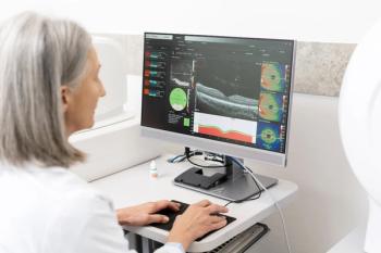
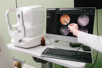
The superficial location of the glands allows for imaging of the glands with transillumination at the slit lamp and with meibography. Due to this growing area of understanding, new technologies make meibography more affordable and both user and patient friendly.
Most commonly used devices for meibography are the Oculus Keratograph 5M, TearScience LipiView II, and a new portable version TearScience LipiScan. The images captured thru meibography not only serve as a great diagnostic for the practitioner but also a valuable tool in educating patients to MGD.
Related:
Classification of gland expression
MGD is diagnosed based on expression of the meibomian glands and classified into two major types based on the meibomian secretion:
• Low delivery states, the most frequent cause
• High delivery states (Figure 3)
Low delivery or obstructive MGD is caused primarily by terminal duct blockage, often from hyperkeratinization of the ductal epithelium, keratinized cell debris, and increased meibum viscosity.6 Cicatricial changes can be noted with obstructive MGD.
The process of low delivery is influenced by both endogenous factors (age, sex, hormones) and exogenous factors (contact lens wear, retinoids, systemic disease).
High delivery or hypersecretion and seborrhea are increases in lipid at the lid margin and are primary or secondary in origin as the case of acne rosacea (Figure 3).
Related:
Treating MGD
To have successful patient outcomes, a standardized exam is needed. Before the TFOS MGD Workshop, treatment of MGD was widely underdiagnosed and treatments varied greatly from one practitioner to the next.
The most common treatments included warm compresses with lid hygiene, artificial lubricants, systemic tetracycline,12 and a topical antibiotic or antibiotic/steroid combination eye drop.
One of the goals for the workshop was to look at evidence-based treatments and develop diagnostic testing and preferred practice guidelines. The TFOS MGD Workshop created a sequence of tests to more systematically diagnose MGD (Table 3).
Once a solid diagnosis with staging of MGD is made, we can refer to the treatment recommendations, which provide the practitioner with an evidence-based approach for the management of MGD (Table 4).
Treatments for MGD vary and include topical artificial lubricants, topical lipid supplements, eyelid warming at home and in-office, mechanical lid hygiene, topical and oral antibiotics, omega 3 fatty acids, and demodex mite management.
Studies have shown that lipid-based artificial tear supplements improve tear film stability and signs and symptoms of MGD.13-16 Eyelid warming and lid hygiene was found to be commonly recommended but poorly standardized.17,18
When treating patients with MGD, evaporative stress must be addressed, including hours spent on digital devices, work environments, home environments, medications, and diet. Treatments to improve meibomian gland function, such as LipiFlow were not part of the MGD workshop as it was only FDA approved following the release of the report.
Since the MGD workshop’s publication, there have been significant expansion in education and treatments for patients on how to manage evaporative stress with blink exercises, new oral supplements, the introduction of LipiFlow thermal pulsation.
At the time of the report, demodex mite blepharitis was not found to be a cause of MGD. And in conclusion of the evidence review it was determined that further randomized controlled masked clinical trials of patients with well-defined MGD are needed to determine efficacy across disease severity.19
Many practitioners are still unfamiliar with the work of TFOS. Raising awareness is crucial to the daily management of patients with DED and MGD.
Use the TFOS MGD Workshop report as your how-to guide for diagnosis and treatment of MGD.
Remember that in the early stages of the disease, patients are often asymptomatic; if left untreated, MGD can cause or exacerbate dry eye symptoms. While MGD is chronic and progressive, it can be effectively treated if diagnosed early.
TFOS is working on the sequel to the original Dry Eye WorkShop, DEWS II (http://tfosdewsreport.org/). DEWS II will update the definition, classification, and diagnosis of dry eye disease; assess the etiology, mechanism, distribution, and global impact of dry eye; and address management and therapy. The DEWS II Report will be available in the first half of 2017.
References
1. Lemp MA, Crews LA, Bron AJ, Foulks GN, Sullivan BD. Distribution of aqueous-deficient and evaporative dry eye in a clinic-based patient cohort: a retrospective study. Cornea. 2012 May;31(5):472-8.
2. Greiner JV, Glonek T, Korb DR, Whalen AC, Hebert E, Hearn SL, Esway JE, Leahy CD. Volume of the human and rabbit meibomian gland system. Adv Exp Med Biol. 1998;438:339-343.
3. Meibom H . De Vasis Palpebrarum Novis Epistola Helmestadi: Typis & sumptibus Helmstadt, Germany: Henningi Mulleri; 1666.
4. Bron AJ, Tiffany JM, Gouveia SM, Yokoi N, Voon LW . Functional aspects of the tear film lipid layer. Exp Eye Res. 2004 Mar;78(3):347-360.
5. Rolando M, Papadia M. Diagnosis and management of the lid and ocular surface disorders. In: Asbell P, Lemp M, eds. Dry Eye Disease: The Clinician's Guide to Diagnosis and Treatment. New York: Thieme Medical Publishers; 2006:71.
6. Schaumberg DA, Nichols JJ, Papas EB, Tong L, Uchino M, Nichols KK. The international workshop on meibomian gland dysfunction: report of the subcommittee on the epidemiology of, and associated risk factors for, MGD. Invest Ophthalmol Vis Sci. 2011 Mar 30;52(4):1994-2005.
7. Nicolaides N, Kaitaranta JK, Rawdah TN, Macy JI, Boswell FM 3rd, Smith RE . Meibomian gland studies: comparison of steer and human lipids. Invest Ophthalmol Vis Sci. 1981 Apr;20(4):522-536.
8. Linton RG, Curnow DH, Riley WJ. The meibomian glands: An investigation into the secretion and some aspects of the physiology. Br J Ophthalmol. 1961 Nov;45(11):718-723.
9. Chew CK, Hykin PG, Jansweijer C, Dikstein S, Tiffany JM, Bron AJ. The casual level of meibomian lipids in humans. Curr Eye Res. 1993 Mar;12(3):255-259.
10. Korb DR, Baron DF, Herman JP, Finnemore VM, Exford JM, Hermosa JL, Leahy CD, Glonek T, Greiner JV. Tear film lipid layer thickness as a function of blinking. Cornea. 1994 Jul;13(4):354-359.
11. Korb DR, Henriquez AS. Meibomian gland dysfunction and contact lens intolerance. J Am Optom Assoc. 1980 Mar;51(3):243-251.
12. Lemp MA, Nichols KK. Blepharitis in the United States 2009: a survey-based perspective on prevalence and treatment. Ocul Surf. 2009 Apr;7(2 Suppl):S1-S14
13. Khanal S, Simmons PA, Pearce EI, Day M, Tomlinson A. Effect of artificial tears on tear stress test. Optom Vis Sci. 2008 Aug;85(8):732-739.
14. Khanal S, Tomlinson A, Pearce EI, Simmons PA. Effect of an oil-in-water emulsion on the tear physiology of patients with mild to moderate dry eye. Cornea. 2007 Feb;26(2):175-181.
15. Simmons PA, Vehige JG. Clinical performance of a mid-viscosity artificial tear for dry eye treatment. Cornea. 2007 Apr;26(3):294-302.
16.Wang J, Simmons P, Aquavella J. Dynamic distribution of artificial tears on the ocular surface. Arch Ophthalmol. 2008 May;126(5):619-625.
17.Smith GT, Dart J. External eye disease. In: Jackson TL ed. Moorfields Manual of Ophthalmology. Philadelphia: Mosby Elsevier; Chap 4:2008.
18.Ehler J, Shah ChP. Wills Eye Manual. Philadelphia: Lippincott Williams & Wilkins; 2008.
19. Geerling G, Tauber J, Baudouin C, Goto E, Matsumoto Y, O'Brien T, Rolando M, Tsubota K, Nichols KK. The international workshop on meibomian gland dystfunction: report of the subcomittee on management and treatment of meibomian gland dysfunction. Invest Ophthalmol Vis Sci. 2011 Mar 30;52(4):2050-2064.
Newsletter
Want more insights like this? Subscribe to Optometry Times and get clinical pearls and practice tips delivered straight to your inbox.


