Clinical findings supported by new OCTA technology
Since its FDA clearance, the significant potential of OCTA has been emphasized.

A 67-year-old male who was the victim of blunt trauma to the right eye 50 years earlier attended for a periodic examination. He was new to the area and admitted to a family history of glaucoma.
His medical history was remarkable only for several uneventful surgical procedures. He took no medications except vitamin supplements. A decade ago he had cataract extraction with intraocular lens (IOL) implantation and subsequent YAG-capsulotomy in the right eye.
Previously from Dr. Semes: When diabetes goes from bad to worse
Exam findings
Visual acuity was correctable to 20/20 in each eye. Age-appropriate lens changes were noted in the phakic left eye. The intraocular pressure (IOP) was measured with Goldmann applanation tonometry at 18 mm Hg in each eye.
Dilated fundus evaluation revealed optic discs consistent with myopic refractive correction, normal retinal vasculature in each eye, age-appropriate macular appearance, and post-traumatic retinopathy superior to the right optic nerve head.
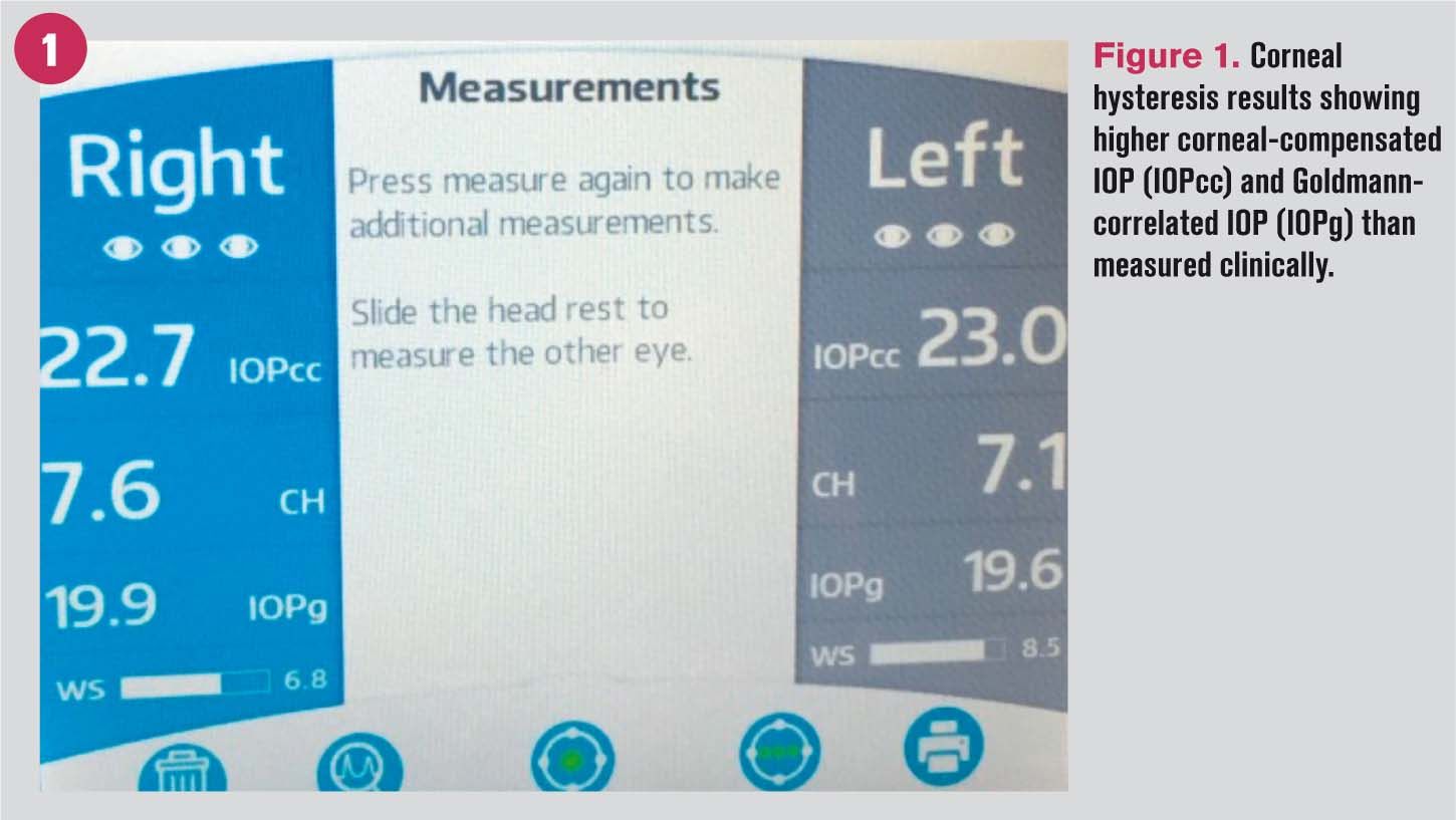
Corneal hysteresis (Figure 1) as well as optical coherence tomography (OCT) and optical coherence tomography angiography (OCTA) were performed to demonstrate changes indicative of glaucoma despite the normal Goldmann applanation tonometry but in view of the positive family history and the patient’s age.

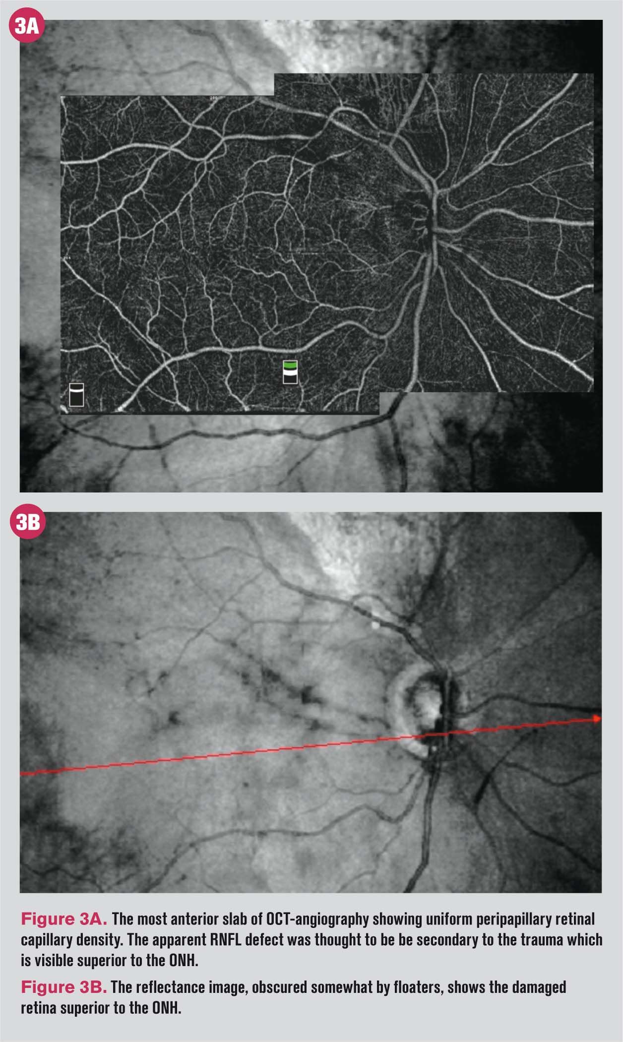
A scan through the area affected by the trauma revealed absence of inner retina (cross-section on OCT-Figures 2A and 2B), and absence of retinal vasculature within both the inner and outer retina on the OCTA images (Figures 3A and 3B). A steep-margined absolute scotoma was demonstrated on threshold visual field testing.
OCTA shows potential
The potential for observing more subtle vascular changes in glaucoma and vascular disorders has been highlighted in the recent literature.1–9


Since its FDA clearance, the significant potential of OCTA has been emphasized. While the present case shows a dramatic loss of retinal vasculature within the remodeled retina, the remainder of circulation, especially to the peripapillary optic disc, appears competent.
References
1. Liu C-H, Wu W-C, Sun M-H, Kao LY, Lee YS, Chen HS. Comparison of the Retinal Microvascular Density Between Open Angle Glaucoma and Nonarteritic Anterior Ischemic Optic Neuropathy. Invest Ophthalmol Vis Sci. 2017 Jul 1;58(9):3350-3356.
2. Bambo MP, Güerri N, Ferrandez B, Cameo B, Fuertes I, Polo V, Garcia-Martin E. Evaluation of the Macular Ganglion Cell-Inner Plexiform Layer and the Circumpapillary Retinal Nerve Fiber Layer in Early to Severe Stages of Glaucoma: Correlation with Central Visual Function and Visual Field Indexes. Ophthalmic Res. 2017;57(4):216-223.
3. Kadomoto S, Muraoka Y, Ooto S, Miwa Y, Iida Y, Suzuma K, Murakami T, Ghashut R, Tsujikawa A, Yoshimura N. Evaluation Of Macular Ischemia In Eyes With Branch Retinal Vein Occlusion: An Optical Coherence Tomography Angiography Study. Retina. 2017 Feb 17.
4. Akil H, Falavarjani KG, Sadda SR, Sadun SA. Optical Coherence Tomography Angiography of the Optic Disc; an Overview. J Ophthalmic Vis Res. 2017 Jan-Mar; 12(1): 98–105.
5. Soares M, Neves C, Marques IP, Pires I, Schwartz C, Costa MA, Santos T, Durbin M, Cunha-Vaz J. Comparison of diabetic retinopathy classification using fluorescein angiography and optical coherence tomography angiography. Br J Ophthalmol. 2017 Jan;101(1):62-68.
6. Ichiyama Y, Minamikawa T, Niwa Y, Ohji M. Capillary Dropout at the Retinal Nerve Fiber Layer Defect in Glaucoma: An Optical Coherence Tomography Angiography Study. J Glaucoma. 2017 Apr;26(4):e142-e145.
7. Esmaili DE. Guidelines for Interpreting OCTA in Retinal Vascular Disease. Retinal Physician. August 2017. Available at: https://www.retinalphysician.com/newsletter/oct-angiography-eupdate/august-2017. Accessed 11/7/17.
8. Gonzalez MA, Shechtman D, Haynie JM, Semes L. Unveiling idiopathic macular telangiectasia: clinical applications of optical coherence tomography angiography. Eur J Ophthalmol. 2017 Jun 26;27(4):e129-e133.
9. Kashani AH, Chen CL, Gahm JK, Zheng F, Richter GM, Rosenfeld PJ, Shi Y, Wang RK. Optical coherence tomography angiography: A comprehensive review of current methods and clinical applications. Prog Retin Eye Res. 2017 Sep;60:66-100.
Newsletter
Want more insights like this? Subscribe to Optometry Times and get clinical pearls and practice tips delivered straight to your inbox.