A closer look at the retina in multiple sclerosis
A 40-year-old black male was evaluated at UAB Eye Care for decreasing vision secondary to multiple sclerosis (MS).

A 40-year-old black male was evaluated at UAB Eye Care for decreasing vision secondary to multiple sclerosis (MS). He had been diagnosed with the disease over 6 years ago and is currently under treatment with Avonex (interferon beta 1a, Biogen Idec). Currently he is a one pack-per-day smoker with a BMI of 27.1. He is an occasional partaker of alcohol (fewer than three drinks per week). Interestingly, he does not report treatment for a bout of optic neuritis. Visual acuity was correctable to 20/30 OD and 20/20 OS with a minimal refractive correction. Pupillary reactivity was sluggish but without differential afferent responses. The anterior segment of each eye was unremarkable and intraocular pressure (IOP) was 14 mm Hg OU (Goldmann applanation).
Fundus evaluation was remarkable for optic atrophy and retinal nerve-fiber layer (RNFL) defects in each eye (Figure 1). In addition, there was an isolated CHRPE lesion observed in each eye. These were considered stable. An optical coherence tomography (OCT) scan was ordered for each eye to investigate quantitatively the RNFL thickness and, more importantly, the ganglion-cell thickness.
MS is a chronic autoimmune central nervous system (CNS) disorder characterized by focal and diffuse inflammation and demyelination as well as neuronal and axonal damage.1 In addition, recent evidence has suggested that average RNFL and macular ganglion-cell thickness is reduced in the disease and that these measures may be useful to establish diagnosis and as markers for progression. These features argue for a diffuse neurodegenerative process.2-6

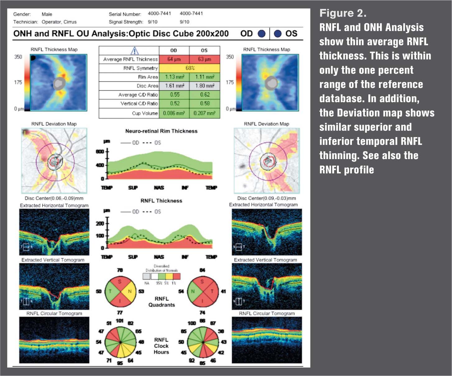
The optic disc cube in each eye is shown in Figure 2. There is good quality signal strength. The average RNFL thickness is 64 and 63 µm OD and OS, respectively. It has been shown that average RNFL thickness is diminished in patients with MS, especially those who have had previous attacks of optic neuritis (ON).6 The optic discs are small, and both the RNFL thickness and deviation maps show RNFL thinning that is consistent with the fundus appearances. The cross-sectional images of the optic discs are incongruent with glaucomatous optic atrophy. The RNFL damage appears to be greater OS, which is the eye with better visual acuity.
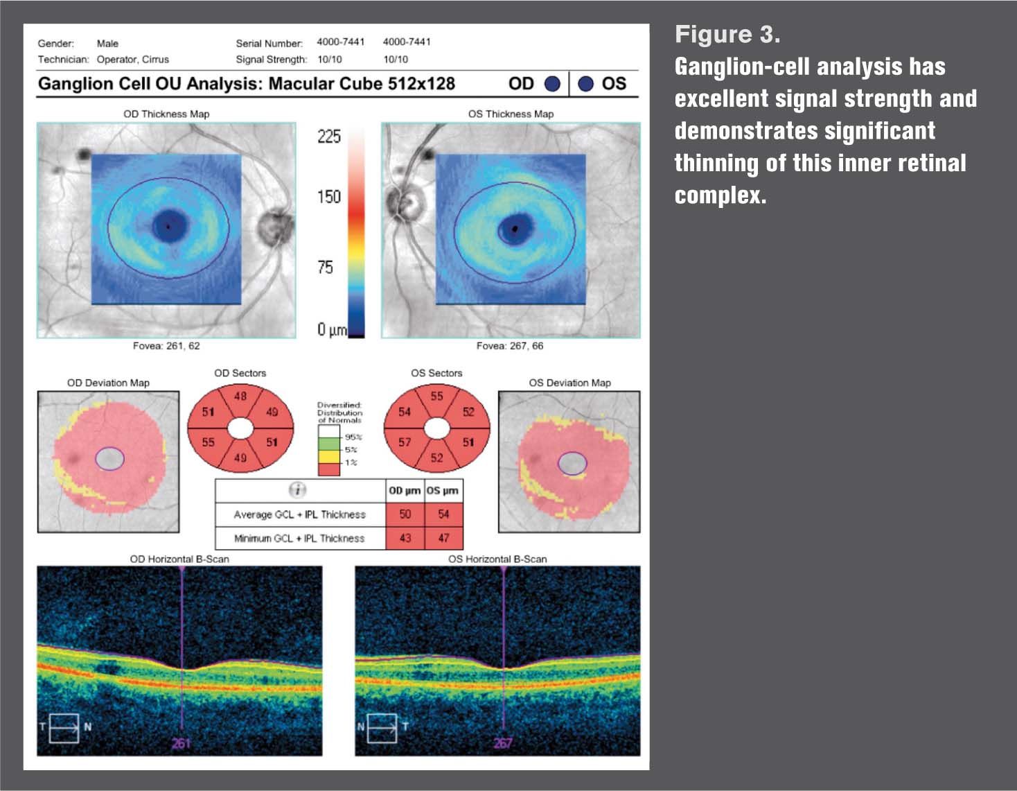
The macular ganglion-cell analysis has excellent signal strength and shows considerable thinning in all sectors as well as grossly on the topographic presentations (Figure 3). It is unlikely that these are artifacts.

Results of two recent studies suggest strongly that serial ganglion-cell loss occurs following acute optic neuritis and offers convincing support that this may be a derivative of optic nerve pathology. What has not been reported is detectable swelling of the inner retina during the acute phase of optic neuritis in MS patients. This thinning was detectable within 3 months of acute optic neuritis and persisted at 6 to 12 months.7,8
What may emerge is the use of OCT and specifically the inner retinal thickness presentations to confirm or even make a diagnosis of MS. Finally, if may be that the inner retina may become the focus for marking subtle changes that indicate progression.

References
1. Compston A, Coles A. Multiple sclerosis. Lancet. 2008 Oct 25;372(9648):1502-17.
2. Bock M, Brandt AU, Dörr J, et al. Patterns of retinal nerve fiber layer loss in multiple sclerosis patients with or without optic neuritis and glaucoma patients. Clin Neurol Neurosurg. 2010 Oct;112(8):647–52.
3. Petzold A, de Boer JF, Schippling S, et al. Optical coherence tomography in multiple sclerosis: a systematic review and meta-analysis. Lancet Neurol. 2010 Sep;9(9):921–32.
4. Syc SB, Saidha S, Newsome SD, et al. Optical coherence tomography segmentation reveals ganglion cell layer pathology after optic neuritis. Brain. 2012 Feb;135(Pt 2):521–33.
5. Albrecht P, Ringelstein M, Müller AK, et al. Degeneration of retinal layers in multiple sclerosis subtypes quantified by optical coherence tomography. Multi Scler. 2012 Oct;18(10):1422-9.
6. Garcia-Martin E, Polo V , Larrosa L-M, et al. Retinal Layer Segmentation in Patients with Multiple Sclerosis Using Spectral Domain Optical Coherence Tomography. Ophthalmology. 2014 Feb;121(2):573-9.
7. Klistorner A, Sriram P, Vootakuru N, et al. Axonal loss of retinal neurons in multiple sclerosis associated with optic radiation lesions. Neurology. 2014 Jun 17; 82(24):2165-72.
8. Ratchford JN, Saidha S, Sotirchos ES, et al. Active MS is associated with accelerated retinal ganglion cell/inner plexiform layer thinning. Neurology. 2013 Jan 1;80(1):47–54.
Newsletter
Want more insights like this? Subscribe to Optometry Times and get clinical pearls and practice tips delivered straight to your inbox.