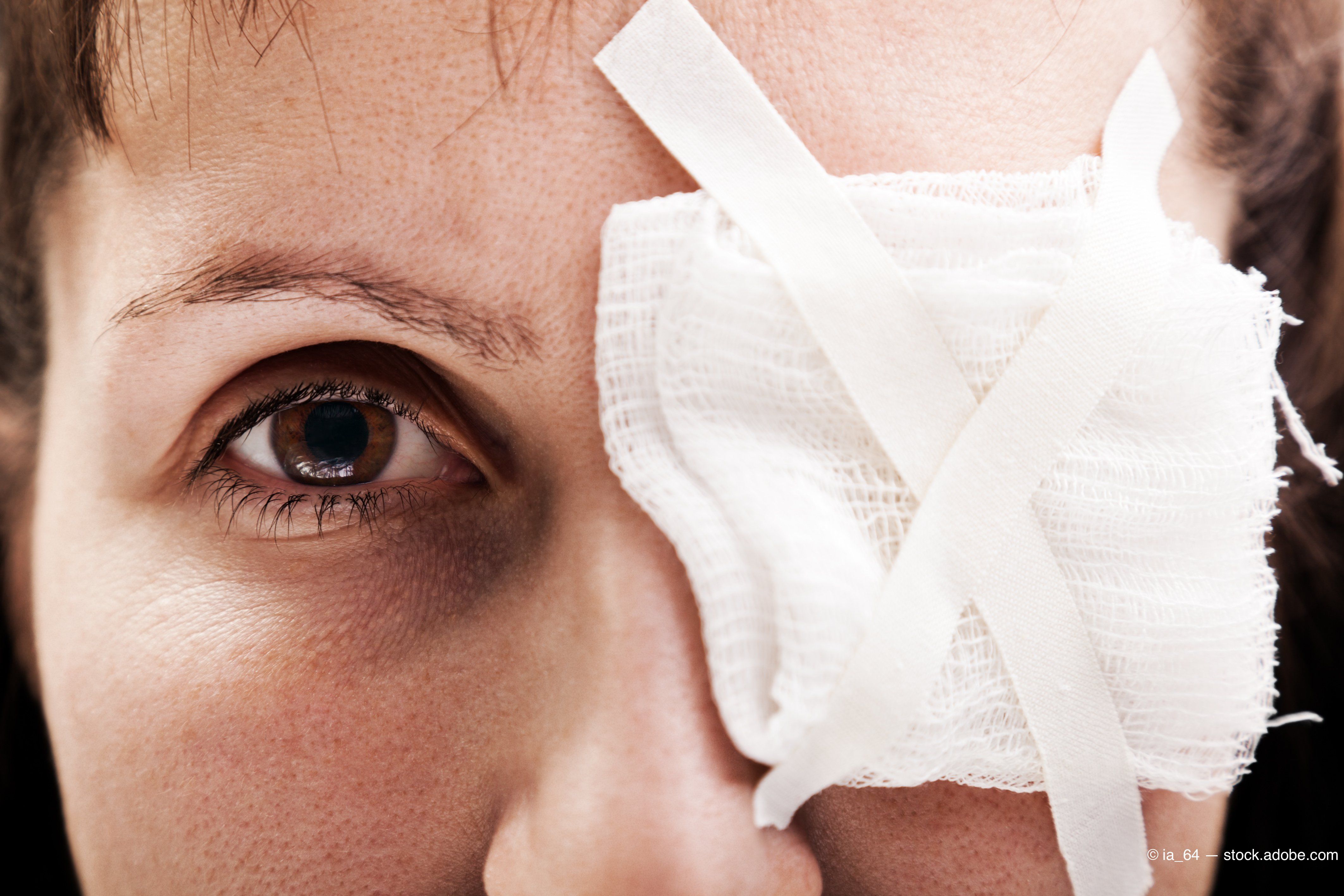
Death of the pressure patch has been slightly exaggerated
Best practice guidelines are just that-guidelines. They’re not religious dogma designed to herd everyone into the same line. We are rightly moving toward more evidence-based medicine, such as increased use of bandage lenses and less pressure patching in the management of corneal abrasions and erosions.


The views expressed here belong to the author. They do not necessarily represent the views of Optometry Times or UBM Medica.
By now, the number of continuing education (CE) lectures I’ve attended is like the cells in a 4+ anterior uveitis-too many to count.
It’s certainly been enough for me to recognize an implicit agreement between lecturer and attendee that constitutes a sacred bond. You know, the one that says, “Lecturers lecture and attendees attend (or at least pretend to), and neither shall harass or attempt to humiliate the other.”
However, one particular CE guru I heard recently apparently didn’t get the memo.
Previously from Dr. Brown: Using the extra eyes within your exam room
It all went down on an early Sunday morning in a dimly lit lecture hall somewhere in the Deep South. Just like church, there were a couple of eager front rowers, but most of the congregation were trying to hide out in the shadows of the back pews.
Sensing that he needed to heat things up, our speaker proceeded to rain down fire and brimstone on our heads.
He warned of the great optometric apocalypse that was nigh upon us and preached on our need to rise up out of our daily ruts, repent our old-fashioned modes of practice, and embrace the glorious gospel of the major paradigm shift.
“How many of you still use a pressure patch?” he yelled into the darkness.
My first reaction was like Admiral Ackbar’s- “It’s a trap!”
But then I thought, OK, what the heck, I’m game, and started to break Rule One of “How to fly low on the radar during CE” by raising my hand.
I didn’t even have a chance to get it fully extended before his eyes latched onto me, apparently the lone sinner in the congregation willing to come forward and confess.
Related: It’s not easy seeing green
“You see?” he shouted, “there’s always at least one! If you don’t use bandage contact lenses (CL), you’re behind the times!”
That’s right: I’d just been “audience shamed.” For a second, I thought he was going to make me don a scarlet “PP.”
Had he actually been willing to listen to what I had to say rather than just using me to make a point, he might have learned that, yes, I do use bandage contact lenses a great deal of the time in patients with corneal abrasions and erosions, as well as in cases of post-corneal foreign body removal.
And yes, I know that most patients prefer them over pressure patches, and they work great.
Except in those cases when they don’t.
Because he wasn’t interested in hearing what those might be, I’ll share a few scenarios in which I might still reach for the tape and eye pads instead of a blister pack-in a non-COPE–approved kind of way, of course.
1. Large, deep epithelial defects
If an abrasion or erosion is small and shallow enough, and the patient isn’t too uncomfortable, I often choose to simply prescribe appropriate topical meds and not use a bandage lens or a patch.
Still, there are defects that are so bad the lid just needs to be immobilized to make the patient comfortable and jump-start the healing process.
Sometimes these come into the office ready-made, and others I create myself when I debride loose strands and flaps of damaged epithelium (Whoa, so that’s what a limbus-to-limbus defect looks like).
Related: UWF: ultra-widefield imaging or ultra-widefield fighting?
After that first big day of re-epithelialization, I might follow with a bandage CL to finish out the period of healing and prevent recurrent erosion.
Yes, I’m aware of the risk of infection in a “warm, moist environment,” but with close follow-up and modern antibiotics, it’s still relatively low.
Exceptions: Contact lens abrasions (and some cases of abrasions from fingernails or vegetative matter).
These patients get a bandage CL or go without cover with an appropriate broad spectrum topical antibiotic and perhaps a short course of a topical nonsteroidal anti-inflammatory drug (NSAID) as an adjunct to cycloplegia for pain control.
2. Lack of patient tolerance and/or contraindications
Surprise, surprise, you aren’t going to be able to apply a CL on all patients.
Some patients can’t stand the idea of having a “foreign object” in their eyes. Syncope on top of everything else, anyone?
For others, the lid swelling and hyper-lacrimation are so severe that attempting to apply a CL is an exercise in futility.
And really, don’t you hesitate just a little in putting a CL on the eye of a patient with severe chronic blepharoconjunctivitis and iffy personal hygiene?
Related: Reliving the joy of your first corneal foreign body
3. No-shows for follow-up
This is a clear and present danger in my practice. There’s usually a better than even chance that one of my patients, for a variety of reasons, is going to be “one and done.”
I’m confident that patients who don’t return for follow-up will reach up, remove a pressure patch, and most likely use the topical meds that I’ve already prescribed for them-at least for a few days.
I’m much less certain of what’s going to happen to a bandage CL in those situations.
It’ll probably fall out on its own, and life will go on.
Or, it could end up as an irritating foreign body in the superior cul-de-sac, or even worse, remain on the cornea for too long and lead to a sight-threatening ulcer.
4. Further mechanical insult
With a pressure patch, the pads and tape can provide a barrier and extra measure of protection against finger or knuckle-to-eye rubbing.
Related: Riding out conjunctivitis like a bad storm
This can be a concern in pediatric and special-needs patients, older patients with dementia, or just absent-minded ones who reach up and start going at it before they realize what they’ve done.
The future of pressure patching
We’ve all learned over the years that less is more. A secure and effective patch doesn’t need a ton of eye pads and tape. No use in sending the patient home looking like King Tut with a six-inch proptosis like we did when we were students.
My broader point is more philosophical than purely clinical, though.
Best practice guidelines are just that-guidelines. They’re not religious dogma designed to herd everyone into the same line.
We are rightly moving toward more evidence-based medicine, such as increased use of bandage lenses and less pressure patching in the management of corneal abrasions and erosions.
But clinical intuition, developed through tens of thousands of patient encounters, enables a clinician to recognize exceptions to “the rule” and offer individualized care to each patient.
Medicine is both art and science. When it comes to reasonable differences of opinion on treatment modalities, perhaps a little more humility would be good for all of us.
I’m betting in a quieter and more reflective setting where he’s away from the bully pulpit and not temporarily lost in the enthusiasm of the moment, my lecturing colleague would say:
Amen.
Newsletter
Want more insights like this? Subscribe to Optometry Times and get clinical pearls and practice tips delivered straight to your inbox.