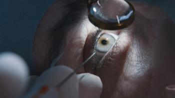
Eye punch leads to choroidal rupture
Significant in her history was blunt trauma to the left eye approximately six years earlier. The nature of this was described as, “…getting a finger poked in my eye.” There were no contributory elements to her medical, family, or social histories.
A 31-year-old female attended to UAB Eye Care for refractive care accompanied by her opposite-sex partner. Her current spectacle lens prescription had been obtained elsewhere; however, her glasses were broken. She reported blurred vision without them. Significant in her history was blunt trauma to the left eye approximately six years earlier. The nature of this was described as, ââ¦getting a finger poked in my eye.â There were no contributory elements to her medical, family, or social histories.
Best-corrected visual acuity was 20/20 in each eye. The anterior segments of each eye were unremarkable. Specifically, there was no evidence of corneal, iris, or lens damage, and intraocular pressure (IOP) was in the statistically normal range. Dilated fundus evaluation of the right eye was unremarkable but revealed the picture shown in Figure 1 for the left eye. This was consistent with a diagnosis of choroidal rupture. On further questioning alone, the patient admitted that the left-eye trauma was secondary to being punched in the eye. This appeared to be more congruent with the fundus damage that was observed.
Optical coherence tomography (OCT) was ordered to better characterize the nature of the choroidal rupture. See Figures 2â4. Additional findings included gonioscopy, which ruled out so-called angle recession. This finding was consistent with the normal IOP finding and lack of optic disc damage.
The patient was advised of the findings and the guarded prognosis following blunt trauma with resulting choroidal rupture. She was advised of the symptoms and risks that can be consequences of choroidal rupture and will be monitored annually unless vision changes intervene.
There is a recent review in International Ophthalmology Clinics on the topic of choroidal rupture.1 Symptoms of choroidal neovascularization would consist of reduced or distorted vision; risks for choroidal neovascularization include older age and macular involvement, which was not present in this case.2
Because of the significant disruption of the outer and inner retina, the patient was asked to return for visual field testing. The purpose of this would be to document the status of retinal sensitivity and follow for any functional changes as well as to make the patient aware of any potential nonseeing areas. The patient refused further testing. Annual surveillance for changes was recommended. In the absence of choroidal neovascularization, primary-care monitoring is appropriate.
References
1. Patel MM, Chee YE, Eliott D. Choroidal rupture: a review. Int Ophthalmol Clin. 2013 Fall; 53(4):69â78. doi: 10.1097/IIO.0b013e31829ced74.
2. Ament CS, Zacks DN, Lane AM, et al. Predictors of visual outcome and choroidal neovascular membrane formation after traumatic choroidal rupture. Arch Ophthalmol. 2006 Jul; 124(7):957â-66.
Newsletter
Want more insights like this? Subscribe to Optometry Times and get clinical pearls and practice tips delivered straight to your inbox.


