Femtosecond advances help ODs, patients
Our patients have numerous choices regarding advanced technology and eye care. Advances range from how patients check in for an appointment to what tools a surgeon uses to dissect tissue. They all have their benefits, and all come at a cost.
Our patients have numerous choices regarding advanced technology and eye care. Advances range from how patients check in for an appointment to what tools a surgeon uses to dissect tissue. They all have their benefits, and all come at a cost.
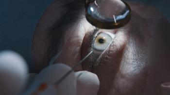
One of the most disruptive, new technologies-both figuratively and literally-is the femtosecond laser (FSL). It is more advanced than robotic surgery in which a computer program tells the laser where to make the cut without a surgeon having to touch an eye.
FSL evolution
FSL is one millionth of a billionth of a second, and the laser operates in the infrared wavelengths ranging from 1,030 nm to 1,053 nm. The laser uses the principle of photoionization vaporizing a small volume of tissue.1
Previously from Dr. Owen:
By lining up these tiny explosions, the laser can create a cleavage plane to separate tissue. The byproduct of this process is mostly carbon-dioxide and water vapor. FSL has the ability to “arrange” the cavitation bubbles in the exact location to separate tissue.2
The first FSL was approved to create a corneal flap for LASIK in 2001. In 2017, the FSL expanded its utility and use to cataract surgery for making incisions, capsulrhexis, liquefying the crystalline lens, and treating astigmatism.
FSL’s role in the cornea has also expanded to removing lenticules for correcting myopia, creating channels for intrastromal rings, making cuts in both the donor and host tissue for penetrating keratoplasty, and lamellar keratoplasty. A mechanical microkeratome may raise the intraocular pressure to as high as 100 mm Hg and advances a diamond blade across the cornea.
Related:
While complications are relatively rare, they can have a dramatic impact on a patient’s clinical outcome. Incomplete flaps, free caps, irregular flaps, and decentered flaps can decrease significantly with the use of FSL.3
FSL creates a planar flap at a more precise depth. Thinner flaps with less deviation from the intended thickness allow the surgeon to comfortably treat higher refractive errors with fewer biomechanical changes.4
The precision of FSL allows for variable flap diameters, increased side-angle cuts, and fewer epithelial defects. These improvements have resulted in few flap displacements, less epithelial in growth, improved corneal sensitivity, and less dry eye.5,6
Cataract surgery and beyond
FSL began its use in cataract surgery in 2008, and it brings increased precision to corneal incisions and the capsulrhexis and allows for the lens to be fragmented with a decrease in phacoemulsification energy.7-9 This decrease in phaco energy results in less endothelial cells loss at one month and in theory may result in a decrease in cystoids macular edema after surgery.10 Limbal relaxing incisions can be made with FSL and are of a precise depth and length, improving refractive outcomes.
FSL LASIK appears to have several advantages over current use of two different lasers. In 2016, the first FSL system (VisuMax, Carl Zeiss) was approved in the United States.
Small-incision lenticule extraction (SMILE) is a procedure where in which a small lenticule of stromal tissue is removed from a corneal incision or flap. The lenticule is of a precise refractive power, resulting in a change in refractive error. SMILE is FDA approved only for the correction of myopia up from -1.00 D to -8.00 D with up to -0.50 D of astigmatism.
Related:
Several theoretical advantages of the SMILE procedure are:
• Less disruption of the corneal nerves, which may result in less dry eye
• Less tissue taking, which may improve biomechanical stability of the cornea and lower induction of higher order aberrations.11-14
Currently, two corneal inlays are approved in the United States, and both require FSL to access the corneal stroma.
The two inlays employ very different strategies to correct presbyopia, but the depth of the inlay is critical to their success. Kamra Inlay (AcuFocus) uses a pin-hole technique to increase depth of focus and increase near vision. A deeper (>220 µm) “pocket” created by FSL yields improved outcomes.15
RainDrop Inlay (ReVision Optics) alters the curvature of the central cornea to create hyperprolate shape to the cornea, resulting in improved near vision. FSL is required to make a pocket at 30 percent of the cornea depth (average 160 µm) to allow for the proper reshaping of the cornea.
Penetrating keratoplasty and lamellar keratoplasty use to create a more precise wound and allow for more elaborate wound configuration-resulting in faster and more predictable wound healing.16,17 Research shows there is less residual astigmatism and a trend toward higher endothelial cell count despite similar final best corrected visual acuity between the two groups.18
The objective of these patterns is to increase the surface area between the donor and the host tissue along with moving the surface junction further from the limbus. ODs aim to decrease the risk of rejection while increasing surface area to promote healing.
Deep lamellar keratoplasty removes the anterior, damaged cornea while leaving a healthy endothelium intact. FSL can precisely remove the anterior tissue while leaving the endothelium untouched.
The laser has been experimentally used to perform sclerotomies to treat glaucoma.19 The precision of FSL allows for an exact incision and a smoother healing.
FSL technology continues to evolve and find more applications within eye care.
Related:
References
1. Kurtz RM, Liu X, Elner VM, Squier JA, Du D, Mourou GA. Photodisruption in the human cornea as a function of laser pulse width. J Refract Surg. 1997 Nov-Dec;13(7):653-8.
2. Chung SH, Mazur E. Surgical applications of femtosecond laser. J Biophotonics. 2009 Oct;2(10):557-72.
3. Durrie DS, Kezirian GM. Femtosecond laser versus mechanical keratome flaps in wavefront-guided laser in situ keratomileusis: prospective contralateral eye study. J Cataract Refract Surg. 2005 Jan;31(1):120-6.
4. Hamilton DR, Johnson RD, Lee N, Bourla N. Differences in the corneal biomechanical effects of surface ablation compared with laser in situ keratomileusis using a microkeratome or femtosecond laser. J Cataract Refract Surg. 2008 Dec;34(12):2049-56.
5. Kezirian GM, Stonecipher KG. Comparison of the IntraLase femtoÃsecond laser and mechanical keratomes for laser in situ keratomileusis. J Cataract Refract Surg. 2004 Apr;30(4):804-11.
6. Tanna M, Schallhorn SC, Hettinger KA. Femtosecond laser versus mechanical microkeratome: a retrospective comparison of visual outcomes at 3 months. J Refract Surg. 2009 Jul;25(7 Suppl):S668-71.
7. Nagy Z, Takacs A, Filkorn T, Sarayba M. Initial clinical evaluation of an intraocular femtosecond laser in cataract surgery. J Refract Surg. 2009 Dec;25(12):1053-60.
8. Abell RG, Kerr NM, Vote BJ. Femtosecond laser-assisted cataract surgery compared with conventional cataract surgery. Clin Experiment Ophthalmol. 2013 Jul;41(5):455-62.
9. Knorz MC. Reduction in mean cumulative dissipated energy following lens liquefaction with an intraocular femtosecond laser. Poster presented at: the American Academy of Ophthalmology Annual Meeting; October 22–25; 2011; Orlando, FL.
10. Chen M, Swinney C, Chen M. Comparing the intraoperative complicaÃtion rate of femtosecond laser-assisted cataract surgery to traditional phacoemulsification. Int J Ophthalmol. 2015; 8(1): 201–203.
11. Li M, Zhou Z, Shen Y, Knorz MC, Gong L, Zhou X. Comparison of corneal sensation between small incision lenticule extraction (SMILE) and femtosecond laser-assisted LASIK for myopia. J Refract Surg. 2014 Feb;30(2):94-100.
12. Denoyer A, Landman E, Trinh L, Faure JF, Auclin F, Baudouin C. Dry eye disease after refractive surgery: comparative outcomes of small incision lenticule extraction versus LASIK. Ophthalmology. 2015 Apr;122(4):669-76.
13. Wang D, Liu M, Chen Y, Zhang X, Xu Y, Wang J, To CH, Liu Q. Differences in the corneal biomechanical changes after SMILE and LASIK. J Refract Surg. 2014 Oct;30(10):702-7.
14. Khalifa MA, Ghoneim A, Shafik Shaheen M, Aly MG, Piñero DP. Comparative analysis of the clinical outcomes of SMILE and wavefront-guided LASIK in low and moderate myopia. J Refract Surg. 2017 May 1;33(5):298-304.
15. Food and Drug Administration. Summary of Safety and Effectiveness Data (SEED). Available at:
16. Lee HP, Zhuang H. Biomechanical study on the edge shapes for penetrating keratoplasty. Comput Methods Biomech Biomed Engin. 2012;15(10):1071-9.
17. Farid M, Steinert RF, Gaster RN, Chamberlain W, Lin A. Comparison of penetrating keratoplasty performed with a femtosecond laser zig-zag incision versus conventional blade trephination. Ophthalmology. 2009 Sep;116(9):1638-43.
18. Chamberlain WD, Rush SW, Mathers WD, Cabezas M, Fraunfelder FW. Comparison of femtosecond laser-assisted keratoplasty versus conventional penetrating keratoplasty. Ophthalmology. 2011 Mar;118(3):486-91.
19. Jin L, Jiang F, Dai N, Peng J, Hu M, He S, Fang K, Yang X. Sclerectomy with nanojoule energy level per pulse by femtosecond fiber laser in vitro. Opt Express. 2015 Aug 24;23(17):22012-23.
Newsletter
Want more insights like this? Subscribe to Optometry Times and get clinical pearls and practice tips delivered straight to your inbox.


