Focus on amblyopia, thyroid eye disease, and papilledema
Clinical pearls on amblyopia, thyroid eye disease, and papilledema treatments from SECO 2022.
Neuro-optometry is an expanding field, with more optometrists becoming more involved in diagnosis and management of some serious commonly presenting neurology-based diseases. Pilar Vergara, DOO, FCOVD, who is in practice in Albacete, Spain, Richard Castillo, OD, DO, from Tahlequah, OK, and Kelsey Moody Mileski, OD, FAAO, from the Marietta Eye Clinic, Marietta, GA, provided their take on these diseases.
Amblyopia: a paradigm shift
Pilar Vergara, DOO, FCOVD, described a new treatment paradigm for managing anisometropic amblyopia. She and colleague Robert Sanet, OD, developed a new protocol to sidestep the shortcomings of the current standard treatment, i.e., patching.
These drawbacks include resistance to wearing the patch, difficulty in school and play because of needing to use the amblyopic eye to process information and perform, disappointing outcomes with long treatment, long-term psychological effects on children and parents resulting from patching, and difficulty achieving 20/20 vision and maximal stereopsis, Dr. Vergara said.
In contrast to the old treatment protocol, which states that the main problem is reduced visual acuity, patching is the optimal treatment, and the optimal prescription is based on most plus to the best visual acuity, Drs. Sanet and Vergara have proposed a new neuroscience-based treatment for amblyopia.
And, indeed, they believe reduced vision is not the problem but only a symptom of the underlying binocular problem.
This thinking represents a paradigm shift.
They described their keys to success as follows: the timing and sequence of each step is crucial; the lens prescription is based on achieving maximal binocularity not maximal visual acuity; unequal adds are frequently prescribed; monocular fixation in a binocular field; optometric phototherapy; use of graded occlusion; and optometric vision therapy.
In cases that are less complicated, that is, defined as a best-corrected visual acuity (BCVA) of 20/50 or better and anisometropia less than 6 diopters (D), using the new protocol, 80% of patients can achieve 20/20 vision and 20 arc seconds of stereopsis without needing any patching over 12 to 15 weeks.
In more complicated cases, defined as a BCVA of 20/50 to 20/200 and anisometropia of up to 8 D or more, most patients can achieve 20/20 to 20/25 vision and 20 to 30 arc seconds of stereopsis without any patching in a maximum of 24 weeks.
Drs. Sanet and Vergara can explain the neuroscience research behind their new model and the steps required to achieve better clinical outcomes. They have designed 2 courses that are currently available: A New Amblyopia Treatment Paradigm and No Tears Amblyopia Treatment-Quicker and Better Results without Patching.
Thyroid eye disease (TED): it takes a village
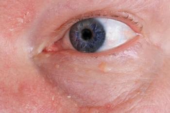
Dr. Castillo described the current knowledge about TED, which manifests most commonly as Graves’ disease (hyperthyroidism, however, also may develop in patients who are hypothyroid and euthyroid (normal hormone levels). The disease is complex, and patients can benefit from the expertise of a number of specialists and subspecialists.
The complexity of this disease is evidenced by the numerous attempts to classify its many manifestations. Presently there are no less than 5 classification schemes. Type I includes cases with minimal inflammation and restrictive myopathy, and type II moderate to significant inflammation and restrictive myopathy.
NO-SPECS includes 7 classes of TED ranging from class 0 with no signs of symptoms to class 6 with sight loss and optic nerve involvement.
The VISA classification, i.e., Vision, Inflammation, Strabismus, and Appearance, defines the vision, inflammation and congestion based on documented changes in inflammation rather than the absolute value, strabismus and motility with ductions and alignments measured, and appearance and exposure. A score of 5 or greater when using this classification indicates active disease or progression for which steroids should be considered.
The EUGODO classification defines mild disease as eyelid swelling and retraction and proptosis; moderate-to-severe disease as active disease that includes diplopia, proptosis of greater than 25 mm, and extraocular muscle dysfunction.
The classical clinical features of thyroid eye disease (Grave’s or hyperthyroid variant) include goiter, exophthalmos, and pretibial myxedema, Dr. Castillo explained.
The risk factors for developing ocular manifestations include increased age at onset, duration of hyperthyroidism, and smoking. These manifestations include eyelid retraction/proptosis, chemosis/conjunctival hyperemia, periorbital edema, diplopia, keratitis/exposure keratopathy, and compressive optic neuropathy.
Examination may reveal eyelid signs that include a staring appearance, lid lag on downgaze, lid retraction, incomplete and infrequent blinking, lower lid edema, and lower lid lag on upgaze.
The standard screening tests for Graves’ disease are thyroid stimulating hormone and free T4. The conventional treatments include beta-blockers, thioamide, radioiodine ablation; surgical approaches are thyroidectomy and TED-specific surgeries, i.e., orbital decompression, strabismus, lid lengthening, blepharoplasty; and most recently teprotumumab infusion (Tepezza, Horizon Therapeutics).
The clinicians involved in treatment include endocrinologists or internists, oculoplastic surgeons, strabismus surgeons, and neuro-ophthalmologists.
“Thyroid eye disease, whether due to the hyperthyroid, hypothyroid, or euthyroid state can be viewed as an immunologic derangement, and understanding the immunopathology involved helps explain the ocular manifestations of TED,” Dr. Castillo said.
Papilledema vs. pseudopapilledema: differentiating the two
Papilledema is a medical emergency and patients should be referred to the emergency room, according to Dr. Mileski.
The disease is characterized by optic disc swelling secondary to elevated intracranial pressure (ICP) that is bilateral in most cases. Elevated ICP causes headache, nausea, vomiting, diplopia, pulsatile tinnitus, and transient visual obscurations.
Papilledema can result from the presence of an intracranial mass, cerebral hemorrhage, meningitis, hydrocephalus, spinal cord lesions, obstruction of venous drainage, and idiopathic intracranial hypertension (IIH). The presentations of these are similar, but all must be excluded from the differential diagnosis before IIH is diagnosed, she advised.
Patients with IIH have normal vision and color vision, possibly diplopia, an enlarged blind spot, and elevated retinal nerve fiber layer and neuroretinal rim on optic nerve optical coherence tomography (OCT). On dilated examination, the optic nerve appears elevated and swollen. Vessel obscuration, Paton’s lines, and hemorrhages may be present.
In contrast, pseudopapilledema is characterized by the absence of an optic disc cup, a small disc, vessel trifurcation, drusen, and spontaneous venous pulsation.
OCT can be useful for differentiating the two diseases by visualizing drusen and peripapillary wrinkles. Fundus autofluorescence (FAF) can also show buried optic disc drusen.
However, the future is promising for diagnosing the two conditions as a result of the use of deep learning systems.
The treatment of papilledema depends on its etiology, that is, a mass or Chiari malformation is addressed surgically, a cerebral venous thrombosis by treating the underlying cause with anticoagulants, and IIH with diet, weight loss, and pharmacologic or surgical therapy.
A new observation about papilledema arising from IIH is that the findings suggestive of IIH on magnetic resonance imaging (MRI) can be the same as in those without IIH. Another new observation is that the prevalence rate of papilledema increased from 2.8% in patients with at least 1 sign on MRI to up to 40% when more signs of IIH were present on imaging
It is also important to remember that anemia can worsen papilledema from IIH. In addition to modifying diet and weight loss, the underlying anemia must also be addressed. The typical treatment of papilledema is acetazolamide (500 mg twice daily and titrated upward to 2 grams daily). Once papilledema is improving, this dosage can be decreased. Optic nerve sheath fenestration may be reserved for severe cases of vision loss.
Dr. Mileski’s clinical pearls include the following:
- Papilledema is a medical emergency
- Symptoms, clinical examinations, OCT, and visual fields all should be used to differentiate papilledema from pseudopapilledema
• Physicians should conduct an urgent outpatient work-up in the event of uncertainty.
• Artificial intelligence may be helpful in the future.
- IIH is a diagnosis that requires needs normal MRI, magnetic resonance venography, and lumbar puncture
- IIH without papilledema is possible but rare
- Patients with IIH should be asked about anemia
“Clinical tools can help differentiate papilledema from pseudopapilledema. If papilledema is suspected, these patients should be sent to the emergency room for proper diagnosis and treatment. Patients need to have normal neuro-imaging (MRI, MRV) and elevated opening pressure for a diagnosis of IIH. These patients should also be tested for anemia as it can exacerbate their condition,” Dr. Mileski said.
Newsletter
Want more insights like this? Subscribe to Optometry Times and get clinical pearls and practice tips delivered straight to your inbox.


