Know what common glaucoma mistakes to avoid
Misdiagnosing neuropathy and over-reliance on OCT imaging results are two examples
Because glaucoma is one of the most prevalent eye diseases in the world, ODs are familiar with best practices for glaucoma management.
But do they really understand glaucoma care as well as they think?
As it turns out, maybe not.
That is according to Joseph Sowka, OD, FAAO, at Nova Southeastern University College of Optometry at the American Academy of Optometry 2019 annual meeting in Orlando.
In particular, optometrists should be aware of several pervasive mistakes in glaucoma management in order to deliver better care to their patients.
Misdiagnosing non-arteritic anterior ischemic optic neuropathy
More than anything else, ODs should avoid diagnosing non-arteritic anterior ischemic optic neuropathy (NAAION) in glaucoma patients.
“It just doesn’t happen,” Dr. Sowka says.
The danger lies in the fact that there is no clinical test to conclusively diagnose this condition, making it a common diagnosis of convenience.
A key differentiator for ODs to keep in mind is that glaucoma is a disease of cupping, while NAAION is a disease of non-cupping.
Over-reliance on OCT
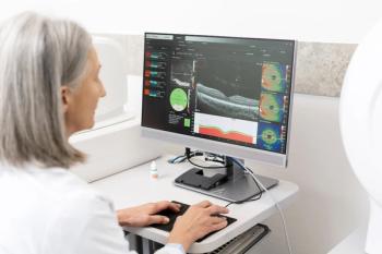
Optical coherence tomography (OCT) is an indispensable tool for diagnosing glaucoma, but a common pitfall inexperienced providers face is overreliance on this imaging.
“The overuse and overemphasis on imaging technology to the exclusion of the other clinical findings and assessment of risk factors is going to put yourself and the patient in some degree of peril,” Dr. Sowka says.
In particular, imaging concerns such as signal quality, blink rates, or segmentation errors can produce erroneous data.
So how much confidence should ODs have in their OCTs for diagnosing glaucoma?
According to Dr. Sowka, optometrists should trust their initial evaluations while factoring in the imaging results. In other words, they should ask themselves how certain they were the patient had glaucoma prior to looking at the OCT imaging results.
“The OCT should not completely change your opinion or your paradigm,” he says.
Thinking that glaucoma causes disc pallor
Another common pitfall optometrists face is the belief that glaucoma causes disc pallor.
As a rule, pallor, in excess of cupping, indicates something other than, or in addition to, glaucoma. Optometrists should assess the physiology of the disc to find clues about which conditions may be present.
“Nothing notches a nerve like glaucoma,” Dr. Sowka says.
Tools such as visual fields used in conjunction with nerve fiber analyses, medical histories, and physical assessments are critical for analyzing these conditions.
“We still need to perform fields in the age of imaging because sometimes it is not glaucoma,” he says.
Mismanagement of red and green diseases
When a disease state masquerades as another condition, it is common for testing values or imaging to fall outside of expected values.
But just because a value falls outside of a standard range, that does not necessarily mean a problem exists. This phenomenon is known as red disease.
“This is a new clinical non-entity,” Dr. Sowka says.
Conversely, there may be glaucomatous processes in a patient masquerading as non-disease. This is known as green disease.
Both of these outcomes tend to increase in prevalence when optometrists place too much trust in imaging tools and machines to make diagnoses. Often, the data inputs that make these decisions may be fallacious.
“Don’t make clinical decisions based on bad data,” Dr. Sowka says.
Newsletter
Want more insights like this? Subscribe to Optometry Times and get clinical pearls and practice tips delivered straight to your inbox.


