Long COVID affects cornea, new study results show
CCM detects small losses of corneal nerve fiber and increases in the number of dendritic cells on the cornea to help identify long COVID
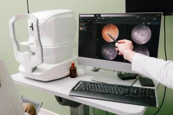
Results from a new study reveal nerve fiber loss and increased dendritic cells (DC) on the cornea may help to identify long COVID. These cells can be found with confocal microscopy (CCM).
About 1 in 10 people who get COVID will develop long COVID, which is defined as symptoms lasting longer than 12 weeks after infection and cannot be explained by another cause.1
Confocal microscopy
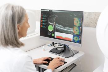
Researchers used a high-resolution imaging CCM to detect small losses of corneal nerve fiber and increases in the number of dendritic cells (DC) on the cornea to help identify long COVID. CCM is commonly used to diagnose various diseases caused by nerve damage, including diabetes, sclerosis, and fibromyalgia.
Senior author Rayaz Malik, MBChB, PhD, with the Department of Medicine, Weill Cornell Medicine Qatar, says most large ophthalmic centers worldwide will have equipment with access to CCM technology, but most academic centers are not likely to be able to offer it due to their size.1
Study results
After being treated and recovering from COVID-19, study participants completed a follow-up survey to see if they had symptoms similar to long COVID. Results showed a strong correlation between those who experienced long COVID and corneal nerve damage.
Neurological symptoms appeared at 4 and 12 weeks in 22 out of 40 (55%) patients and 13 out of 29 (45%) patients, respectively.1
"COVID has affected so many parts of the body, it's no surprise that it affects the eye,” says Angie Wen, MD, assistant professor of ophthalmology, cornea, cataract, and refractive surgery at New York Eye & Ear Infirmary of Mount Sinai.
Checking the cornea through CCM could serve as another piece of information to support a long COVID diagnosis for people who have had, for instance, pulmonary symptoms and unexplained neuropathic pain, says Wen. "But it seems we're unable to draw that conclusion yet from these results,” she says.
Study limitations
The study is limited by scope and number of participants. Additionally, participants were asked to define the severity of their neurological symptoms in a questionnaire. The subjective nature of responses may have also skewed outcomes.
Reference
1. Bitirgen G, Korkmaz C, Zamani A, et al. Corneal confocal microscopy identifies corneal nerve fibre loss and increased dendritic cells in patients with long COVID. Br J Ophthalmol. 2021.:bjophthalmol-2021-319450. doi:10.1136/bjophthalmol-2021-319450
Newsletter
Want more insights like this? Subscribe to Optometry Times and get clinical pearls and practice tips delivered straight to your inbox.


