Managing a partial-thickness laceration
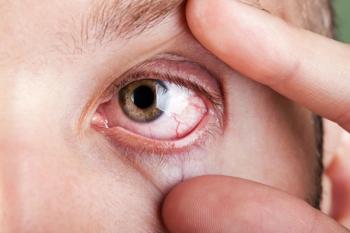
A 28-year-old white male presented with the complaint of a scratched right eye. He reported that earlier that day, he had been working on a construction project and was hammering a piece of plastic when the plastic splintered and hit him in the right eye.
A 28-year-old white male presented with the complaint of a scratched right eye. He reported that earlier that day, he had been working on a construction project and was hammering a piece of plastic when the plastic splintered and hit him in the right eye. He described that initially there was a stabbing pain, and then the eye began watering profusely. A Seidel test was negative, and he was prescribed topical Ciloxan (ciprofloxacin, Bayer) to be used every hour while awake.
Follow-up the next day
The patient returned the following morning for a scheduled follow-up visit. He complained of severe watering, photophobia, foreign body sensation, and redness OD. Uncorrected visual acuity was 20/20- OD. There was a severe ptosis OD, Grade 2-3+ conjunctival injection, and a 4 mm linear opacity directly under the visual axis. Surrounding the linear opacity was epithelial and stromal edema. The anterior chamber was fully formed. The slit lamp appearance is shown in Figure 1.
There was also a 3+ anterior chamber reaction as shown in Figures 2 and 3. For the follow-up, fluorescein was instilled to inspect the linear opacity. The appearance of the fluorescein staining is demonstrated in Figure 4.
The opacity had re-epithelialized overnight and had both positive and negative stain. A repeat Seidel test was negative. The exterior lid was evaluated and then everted to inspect for a retained conjunctival foreign body. Papillae (Grade 2+) were present, but there was no foreign body.
Anterior segment optical coherence tomography (ASOCT) was then performed at the site of the wound. A line scan at the deepest part of the wound is shown in Figure 5.
Upon further investigation, the pupil was sluggish but reacted to light. Intraocular pressure (IOP) by applanation tonometry was 11 mm Hg. A careful dilated fundoscopic evaluation showed no sign of a retained intraocular foreign body.
Managing a laceration
The patient was diagnosed with a partial-thickness corneal laceration and traumatic iritis and prescribed the following: decrease ciprofloxacin (Cipro, Bayer) to qid OD; prednisolone acetate 1% (Pred Forte, Allergan) q2h OD; Muro (sodium chloride hypertonicity ophthalmic ointment, Bausch + Lomb) 128 qid OD; homatropine 5% (Isopto Homatropine, Alcon) bid OD; and he was instructed to purchase a shield for sleeping at night. A return visit was scheduled for three days.
The patient returned for a follow-up visit three days later. He reported that his eye felt better. The foreign body sensation had resolved, although he said his eye did not feel that it was back to normal. The redness had improved significantly and vision was unchanged. He was taking the ciprofloxacin qid OD, prednisolone acetate q2h OD, and Muro 128 qid OD, but he did not fill the Rx for the prescribed homatropine 5% bid. Uncorrected visual acuity was 20/20-1 OD.
A repeat line scan ASOCT was performed along the axis of the linear laceration and is shown in Figure 6. The line scan along the axis of the linear laceration revealed a highly reflective object within the laceration. Figure 7 demonstrates the slit lamp appearance of that retained foreign body.
Biomicroscopy showed resolution of the ptosis OD, resolution of the palpebral conjunctival papillae, trace bulbar conjunctival injection, a linear corneal laceration measuring 4 mm with complete re-epithelialization (using NaFl stain) and minimal corneal edema surrounding, a retained foreign body within the laceration, 2-3+ cells and flare in the anterior chamber, and IOP of 14 mm Hg.
A higher magnification image of the retained foreign body is shown in Figure 8.
Upon review of the initial images taken, the retained foreign body was present but difficult to identify though the corneal edema and roughened epithelial surface.
The continuation of the management plan was for the patient to fill the homatropine 5% prescription, discontinue the topical ciprofloxacin, continue the Pred Forte at q2h OD, and continue Muro 128 tid OD. The patient was then asked to find the manufacturer of the plastic electrical box and then determine if the plastic used in its manufacture is a stable or leeching material. The patient was scheduled to return to clinic in seven days for further follow-up.
Later that day, he e-mailed a report of the plastic used in the box that shattered and entered the cornea. The box was made from polyvinyl chloride (PVC).
Discussion
The major concern in any corneal laceration is the possibility of full-thickness penetration. A full-thickness laceration, or ruptured globe injury, will often allow aqueous humor to leak, often described by patients as severe tearing. Despite the elegance of the Seidel test, a full-thickness laceration can be present even with a negative Seidel test,1 and the test is often difficult to interpret because of the associated tearing. Other signs in a ruptured globe may include a flat chamber, air bubbles in the anterior chamber, and reduced visual acuity. If a ruptured globe is suspected and ASOCT is not available, applanation tonometry should not be performed. Ruptured globe injuries require immediate surgical management. A partial-thickness corneal laceration is managed medically. A scar will result when Bowman’s membrane is penetrated.
With a retained foreign body, one must determine the composition of the invasive material. Metallic foreign bodies must be removed either at the biomicroscope or surgically in the operating room. However, hard, non-leeching plastics may be allowed to reside within the cornea. Recall that the genesis of hard contact lens development was spurred by the observation that retained foreign bodies of shards of shattered PMMA plastic cockpit canopies in World War II fighter pilots did not react with the corneal tissue.
In this case, the plastic was made from PVC. The PVC trade group states, “PVC is safe, chemically stable, inert, extremely versatile and easily fabricated. Medical products made from PVC are usable inside the body, easy to sterilize, and simple to assemble into products that do not crack or leak.”2
References
1. Kah TA, Salowi MA, Tagal JM, et al. Occult open globe injury in a patient with corneal foreign body: a case report. Cornea. 2009 Dec;28(10):1164-6.
2. PVC. PVC for Health. Available at:
3. Parver LM, Dannenberg AL, Blacklow B, et al. Characteristics and causes of penetrating eye injuries reported to the National Eye Trauma System Registry, 1985-91. Public Health Rep. 1993 Sep-Oct;108(5):625-32.
Newsletter
Want more insights like this? Subscribe to Optometry Times and get clinical pearls and practice tips delivered straight to your inbox.


