New device enables imaging of meibomian gland structures
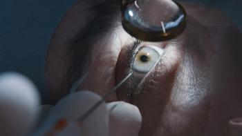
A new multipurpose diagnostic instrument, featuring an infrared illumination system, is a useful tool for imaging meibomian gland structures in the upper and lower lids.
Key Points
"Recognition that meibomian gland dysfunction is the major cause of evaporative dry eye has focused attention on the importance of examining the meibomian glands. It is an aspect of the clinical evaluation, however, that is often overlooked. Slit-lamp evaluation is sufficient only to evaluate the lid and lid margin, while infrared meibography enables visualization of meibomian gland structure," said Dr. Srinivasan, research assistant professor, University of Waterloo, School of Optometry, Waterloo, Ontario.
"The platform we used is a noncontact, user- and patient-friendly instrument that allows for noninvasive, rapid evaluation of meibomian gland structure along with multiple other diagnostic functions," she explained.
"All these applications are useful in clinical practice, and capturing of meibography images could be done without making any modifications or adaptations to the instrument. Visualization of the glands and image capture was completed in less than 1 minute," noted Dr. Srinivasan.
Imaging structure, changes
The study she reported aimed to examine the ability of the device to image the meibomian gland structures and to investigate possible morphological changes in the eyelids of young adults. Dr. Srinivasan and colleagues enrolled 37 participants (13 men and 24 women), including 24 contact lens wearers. Subjects ranged in age from 19 to 45 years, and the mean age of the contact lens wearers and control group was similar (28 and 25 years, respectively).
"There are some disadvantages in using this device for studying meibomian gland structure. As it was not specifically designed for meibography, maneuvering the head of the instrument for this task is a little difficult, and it has limited magnification. Therefore, we are planning to use confocal microscopy to further assess some of the changes we observed," said Dr. Srinivasan.
Gland dropout was assessed using a four-point scoring system reported by Arita et al. (Ophthalmology. 2008 May;115(5):911-915) in which 0 = no gland loss, 2 = >1% to 33% loss, 2 = 33% to 67% loss, and 3 = >67% loss. A "meiboscore" was calculated for each subject by summing the upper and lower lid scores, and the data were averaged for each study group.
Meiboscores ranged from 0 to 5 in the contact lens wearers and from 0 to 2 in the controls. Mean meiboscores were similar in the left and right eyes of the contact lens wearers, and for a the right eye, the mean score was significantly higher in the contact lens wearers compared with the controls. However, for the left eye, there was only a trend for the mean meiboscore to be significantly higher in the contact lens wearers compared with the controls.
The contact lens wearers had a significantly higher (worse) OSDI score than the controls (16 versus 7.5). There was a statistically significant, positive correlation between the OSDI score and meiboscores in the contact lens wearers. In this small study, possible correlations between lens type and meiboscores were not investigated.
"Interestingly, tortuosity was observed only on the upper lids in nine study participants, of which seven were female contact lens wearers and two were male controls. More data and further study are warranted to try to understand this finding, and we will be investigating correlations between tortuosity and meibum quality. In addition, we will be using confocal microscopy to evaluate acinar density and to examine the white patches, which we believe are aging changes," Dr. Srinivasan said.
Moving forward, the researchers have also begun to use digital image analysis for calculating percentage of gland loss in order to overcome interobserver variability.
FYI
Sruthi Srinivasan, PhD, BSOptom, FAAOE-mail:
Oculus supplied the instrument for the study and Allergan provided an educational grant. Dr. Srinivasan has no direct financial interest to report.
Newsletter
Want more insights like this? Subscribe to Optometry Times and get clinical pearls and practice tips delivered straight to your inbox.


