
News|Articles|December 18, 2024
New study indicates DNA damage as key factor in AMD
Author(s)Jordana Joy, Associate Editor
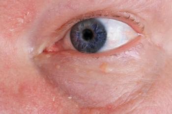
Mice with reduced levels of ERCC1-XPF in the study presented abnormal optokinetic and electroretinogram responses consistent with photoreceptor dysfunction and visual impairment.
Advertisement
A new study’s research team, co-led by the University of California Irvine, has discovered that accumulated DNA damage in the retina is a key contributor to age-related macular degeneration (AMD).1,2 In their study, which was recently published in Aging Cell, authors also found that targeting specific retinal cell types may lead to treatments that can slow or stop progression, according to a news release.
“Our findings highlight the critical role DNA damage repair plays in maintaining retina health for good vision,” said co-corresponding author and UC Irvine associate professor of physiology and biophysics Dorota Skowronska-Krawczyk, PhD. “Because age is the strongest risk fact for AMD, gaining deeper insights into the underlying biology of aging in the eye is essential for developing effective therapies.”
The research comprised of comparing a mouse model with reduced levels of ERCC1-XPF (Ercc1−/Δ), a DNA repair enzyme, aged 2 and 4 months old, with 2 control groups: young, aged-matched mice and 30 month old, naturally aging mice.2 The neural retina and retinal pigment epithelium (RPE) from Ercc1−/Δ mice were compared to the age-matched and older mice by means of optokinetic response measurement, electroretinogram, RNA isolation and qPCR, hematoxylin and eosin staining, color fundus photography, fluorescent immunostaining, RNAscope in-situ hybridization, staining of RPE flat mount and retinal wholemount, measuring oxygen consumption and glycolysis in RPE, mitochondria and mitophagy staining of RPE cells, C12FDG staining, and immunoblotting.2
By 3 months of age, the Ercc1−/Δ mice had presented abnormal optokinetic and electroretinogram responses consistent with photoreceptor dysfunction and visual impairment.1,2 Between ages 3-4 months, Ercc1−/Δ mice also showed signs of visual impairment, structural alterations in the retina, abnormal blood vessel formation, shifts in gene expression and metabolism, and mitochondrial dysfunction in the retinal pigment epithelium.2 According to the release, these changes mirror those seen in a typical human eye when aging.1 “Hence, our study suggests spontaneous endogenous DNA damage promotes the hallmarks of age-related retinal degeneration,” the study authors stated.
“The more we know about how DNA damage contributes to eye diseases like AMD, [the better] we can develop interventions that address the root causes of vision loss. These could include strategies to counteract oxidative stress, enhance DNA repair, or even remove damaged cells before they cause harm,” Skowronska-Krawczyk said. “We plan to investigate which cell types drive age-related changes by selectively impairing DNA mechanisms. Our goal is to advance the development of preventative interventions that significantly reduce the burden of age-related vision loss and improve the quality of life for millions.”
References:
UC Irvine-co-led study finds DNA damage is key factor in age-related macular degeneration. News release. University of California Irvine News. December 3, 2024. Accessed December 12, 2024.
https://news.uci.edu/2024/12/03/uc-irvine-co-led-study-finds-dna-damage-is-key-factor-in-age-related-macular-degeneration/ Narasimhan A, Min SH, Johnson LL, Roehrich H, Cho W, Her TK. The Ercc1−/Δ mouse model of XFE progeroid syndrome undergoes accelerated retinal degeneration. Aging Cell. 2024 Nov. e14419.
https://doi.org/10.1111/acel.14419
Newsletter
Want more insights like this? Subscribe to Optometry Times and get clinical pearls and practice tips delivered straight to your inbox.
Advertisement
Related Content
Advertisement


Latest CME


Advertisement
Advertisement
Trending on Optometry Times - Clinical News & Expert Optometrist Insights
1
Greenland sharks may hold secret to visual preservation
2
Mulling over options for mitigating severe vision loss from diabetes
3
Herpes zoster ophthalmicus in the V2 dermatome post Shingrix vaccination
4
GSLS 2026: Scleral lens troubleshooting with Dr Elise Kramer
5