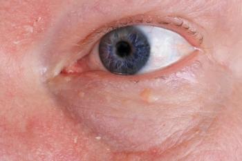
Novel anterior segment and retinal camera is used by NASA astronauts
A novel anterior segment and retinal camera attachment is used to take still pictures and video of the eye. Then, using a computer link, images and video clips can be sent for review anywhere in the world. NASA purchased the device for use at the International Space Station.
Key Points
Sturgeon Bay, WI-A Wisconsin optometrist's invention has taken off-literally. Among the equipment assigned to the space shuttle Discovery was an anterior segment and retinal camera attachment created by Paul Filar, OD.
Dr. Filar conceived the device so that it could go anywhere, which now includes outer space. Astronauts will be able to use the retinal camera attached to an ophthalmoscope to relay pictures to medical professionals on Earth to diagnose any problems with their vision.
The device (Provizion Anterior Segment and Retinal Camera Attachment) is designed to fit on a Welch Allyn PanOptic Ophthalmoscope. The attachment is used to take still pictures and video of the eye, particularly the optic nerve. Then, using a computer link, images and video clips can be sent for review anywhere in the world-or, in this case, to Earth from space.
Dr. Filar said he's accustomed to patients who squirm and blink, making it difficult to study the anterior segment and retina. He wanted a mobile piece of equipment that made it easy to document, enlarge, and study images without inconveniencing patients.
"If I'm in a nursing home and the patient is hunched in a wheelchair, I can take the photo and look at it later," he said. "I needed to be able to photograph, document, and download it to my computer. I wanted to have something in case there was an emergency or bleeding behind the eye.
"All of the other equipment that does that is big and bulky and costs about $40,000," Dr. Filar said. He needed another option for getting eye images back to his office for review. So, Dr. Filar developed his own alternative.
"I like to try and find different solutions to a problem," he said.
The device is a user-friendly, portable, and billable instrument. The camera is encased in a plastic housing, and can take images up to 8 megapixels (software enhanced).
The camera attachment software features options including auto or manual focusing, time and date stamping, zoom, and high-definition video capability. When photography is completed, remove it from the ophthalmoscope and use a USB cable to connect it to any computer.
Thinking in terms of telemedicine, Dr. Filar saw wide applications for his invention. "It could have an application in emergency rooms and other types of care," he said.
Among the markets he envisioned serving were the military, veterinarians, other eye doctors, and missionaries in impoverished nations-but never the space station.
"It makes taking a picture or video affordable," Filar said. "So I knew it was good for taking pictures in remote areas-but the NASA application is not something I ever thought of."
"This all happened very quickly," said Keith A. Favaro of Burlington, WI, whose 20/20 Medical Services equipment distribution company markets Dr. Filar's camera.
Dr. Filar made a sheet-metal prototype and took it to a local plastics manufacturer to see if he could produce it.
Mike Keyser, owner of Key Industrial Plastics, said he was impressed with Filar's invention from the start.
"I get a lot of people that come in with ideas. Most of the time, it's from a large corporation," Keyser said. "This one was designed by a professional who knew what he was doing, and I knew we could make this."
The doctor filed a patent application for the device in February and production started in April.
He and his distributor displayed it at a Wisconsin medical conference in April.
Sales of the device were launched through a Web site,
"We got a phone call from NASA," Dr. Filar said. Shocked and energized, he realized that this was the perfect application. The unexpected call from NASA resulted in a priority order for two devices-one to go up to the International Space Station and the other for training here on Earth. NASA checked the weight to ensure they would be compatible with NASA's in-flight computers. Ten days later, NASA ordered four more.
"The device is scheduled to be left at the International Space Station as a medical tool to allow the flight surgeons and research community insight into potential changes in the eye that may come about as a result of long-duration missions," said William Jeffs, a spokesman for the NASA Johnson Space Center in Houston.
As a sci-fi buff, Dr. Filar is especially excited about being able to contribute something to the space program.
This isn't the first of Dr. Filar's optical inventions, and he said it wouldn't be his last. He created a device to help stroke victims called a bi-nasal occluder, which blocks off part of the vision field to develop brain activity. It was sold to a therapy company 6 years ago.
He is a graduate of St. Norbert College, De Pere, WI, and Pacific University's College of Optometry, Forest Grove, OR. The Wisconsin State Optometric Association and the American Optometric Association named him Wisconsin 2008 Young Optometrist of the Year.
Newsletter
Want more insights like this? Subscribe to Optometry Times and get clinical pearls and practice tips delivered straight to your inbox.


