Optical coherence tomography aids retinal diagnosis, treatment
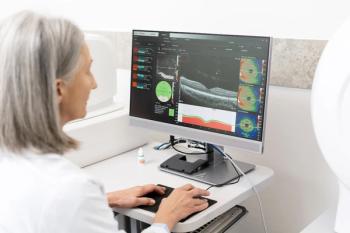
Optical coherence tomography (OCT) can be an effective diagnostic tool that enables optometrists to examine the retina layer by layer. It also can offer quantitative data about the retinal nerve fiber layer. Some doctors believe that the biggest advantage of OCT is what it reveals about the vitreomacular interface.
Key Points
When checking the time on his digital clock, he only could see the first and last numbers, not the middle digit, even though he was staring straight at the clock. After dilating the pupils and examining the macula with slit lamp biomicroscopy, Leo Semes, OD, FAAO, used optical coherence tomography (OCT) to determine whether the retinal pigment epithelium had been compromised, a state that can signal choroidal neovascularization. "Clinically, there was evidence of macular disruption," said Dr. Semes, professor of optometry, University of Alabama at Birmingham (UAB).
The man also was participating in a clinical trial that examined the impact of zeaxanthin on macular function, he added. "With the OCT, we were able to demonstrate a disruption of retinal pigment epithelium (RPE) and confirm neovascularization very early," Dr. Semes said.
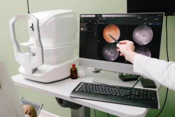
Many optometrists use or have access to OCT, a diagnostic tool that Dr. Semes said allows doctors to "cheat" by examining the retina layer by layer. The instrument's data gathering is akin to in vivo histology. Although some optometrists are using OCT to effectively diagnose and treat disease in patients, others such as Dr. Semes also are exploring creative applications for the technology.
Dr. Semes said he has used OCT for more than 3 years. Although OCT is in its infancy for glaucoma, he said that it is his technology of choice for patients with diabetes or macular degeneration.
Consider a prodigal diabetic patient, aged 49 years, who had mild, nonproliferative retinopathy and good vision. A clinical examination revealed some evidence of swelling that did not qualify as clinically significant macular edema. When interpreting the OCT in consultation with a retina specialist, however, he said, a treatment plan was devised for treating diabetic macular edema that was encroaching on the macula.
Another one of Dr. Semes' patients had macular edema secondary to a branch vein occlusion. Despite several months of treatment using topical non-steroidal anti-inflammatory drugs, structure and function were still disturbed. After an OCT exam, Dr. Semes believed that the patient was a good candidate for anti-vascular endothelial growth factor therapy in an attempt to re-establish the integrity of the retinal vasculature. Following a single injection, the patient's vision returned to 20/25 from 20/70.
The plus factor
Dr. Semes said he believes that the biggest advantage of the OCT is what it reveals about the vitreomacular interface. "For example, it's really elucidated and confirmed the evolution of macular hole formation," he said. "It allows that to be monitored very carefully and has given us a much better idea for when surgical intervention is indicated and whether it has been successful."
OCT also can offer quantitative data about the retinal nerve fiber layer, Dr. Semes said. The thickness of the fiber layer is a parameter that is emerging to distinguish patients with healthy, normal eyes from those with very early glaucoma changes, he added. But when progression occurs is indicated, he said, OCT may suggest the need for alternative interventions.
Meanwhile, his department is conducting a pilot study involving patients who have demonstrated early AMD changes. The study is designed to compare thinning of the RPE with results from a preferential hyperacuity perimeter device (Foresee PHP, SightPath Medical) to see how they correlate with the OCT's structural results. Data collection will be completed by the end of this year.
"Perhaps once we have a better handle on what changes occur based on the data collected, maybe we'll be able to understand how certain eye diseases progress, how they can be treated more successfully," Dr. Semes said. "We're still developing, improving, and evolving."
Newsletter
Want more insights like this? Subscribe to Optometry Times and get clinical pearls and practice tips delivered straight to your inbox.


