Posterior view critical pre-refractive surgery
Examination of the posterior surface of the cornea is critically important when evaluating patients for refractive surgery, one expert notes.

Key Points

While optometrists and ophthalmologists have generally examined the anterior surface of the cornea prior to refractive surgery, in most cases the posterior surface is more important to consider, Dr. Morgenstern said. The technology to examine the posterior surface exists the form of a rotating Scheimpflug camera (Pentacam, Oculus), and a proprietary corneal topographer (Orbscan IIz, Bausch + Lomb), he added.

"There's always risk involved with surgery, it doesn't matter what the surgery is," he added. "Any patient can have an aberrant outcome from LASIK or PRK, but with proper screening, we can maximize the chance of a healthy outcome."
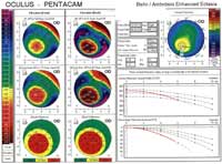
The usefulness of the rotating Scheimpflug camera can be further enhanced with software upgrades. On the Oculus product, for example, Dr. Morgenstern cited an available feature called the Belin/Ambrosio Enhanced Ectasia Display that combines elevation-based mapping and pachymetric corneal evaluation in an all-inclusive display.
"Basically, this feature compares curves and points of elevation on the front and back surface of the cornea to give the doctor a warning if there's a patient who's a bad candidate [for a refractive procedure]. It is like a diagnostic tool in the software. You have to upgrade to it, but it is worth its weight in gold to help you determine good or bad candidates," he said.
He added that the rotating Scheimpflug camera also is an outstanding machine for specialty contact lens practices that work with patients who are hard to fit due to keratoconus.
"It is a very valuable tool for early detection and monitoring of progression of keratoconus," Dr. Morgenstern said. "Because of both this device and the proprietary topographer, keratoconus has gone from being an anterior surface disease to a posterior surface disease. We see the earliest warning signs of keratoconus on the backside of the cornea. By the time it gets to the front surface, it is more advanced. But, in the past, we only had the capability to look at the front surface."
The rotating Scheimpflug camera's densitometry reading also makes it useful for monitoring cataract progression, he said.