Researchers develop eye drops that slow progressive vision loss in mice
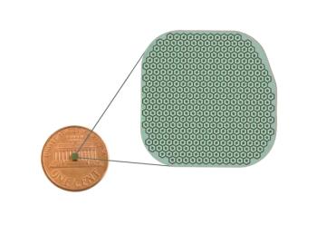
Researchers developed eye drop formulations 17-mer and H105A, both containing a short peptide, to administer to mice in the form of a drop on the eye’s surface.
Eye drops that extend vision in animal models of a group of inherited diseases that lead to progressive vision loss in humans was recently developed by researchers at the National Institutes of Health (NIH).1,2 The researchers, who published a report on the study in Communications Medicine, developed eye drops that contain a small fragment derived from a protein in the body and found in the eye known as pigment epithelium-derive factor (PEDF). It has been found that PEDF helps preserve cells in the retina.
“While not a cure, this study shows that PEDF-based eye drops can slow progression of a variety of degenerative retinal diseases in animals, including various types of retinitis pigmentosa and dry age-related macular degeneration (AMD),” said Patricia Becerra, PhD, chief of NIH’s Section on Protein Structure and Function at the National Eye Institute and senior author of the study, in a news release. “Given these results, we’re excited to begin trials of these eye drops in people.”
In the study, led by Alexandra Bernardo-Colón, researchers used specially bred mice that lose their photoreceptors shortly after birth. Once cell loss begins, the majority of the mice photoreceptors die in a week. Researchers created 2 eye drop formulations, each containing a short peptide, to administer to mice in the form of a drop on the eye’s surface. One of the formulations, 17-mer, contains 17 amino acids found in the active region of PEDF. H105A, the second formulation, is similar to 17-mer but binds more strongly to the PEDF receptor.1
According to the release, when peptides were applied to the mice’s eyes in a 5–10 μl eye drop,2 a high concentration in the retina was found within 60 minutes, slowly decreasing over the next 24 to 48 hours. Through the 1-week period of treatment, mice retained up to 75% of photoreceptors and continued to have strong retinal responses to light. Additionally, both 17-mer and H105A eye drops decreased pro-apoptotic BAX and increased anti-apoptotic BCL2 levels in photoreceptors. When given a placebo, mice had few remaining photoreceptors and little functional vision at the end of the week. Neither peptide formulation caused toxicity or other side effects in the study.1
“For the first time, we show that eye drops containing these short peptides can pass into the eye and have a therapeutic effect on the retina,” said Bernardo-Colón in the release. “Animals given the H105A peptide have dramatically healthier-looking retinas, with no negative side effects.”
Study authors noted that further research is necessary to optimize peptide delivery methods and establish long-term safety and efficacy.1
“The significance and potential impact of our study are grounded on its translational promise as it demonstrates the ability of 17-mer and H105A peptides to stabilize the progression of photoreceptor degeneration in preclinical murine models of human retinal diseases … Not only did the peptides act on preserving photoreceptor morphology but also on improving the light responses of both types of photoreceptors, rods, and cones. These genetically distinct models have common and unique retinopathies, suggesting that the H105A peptide appears to have broad applicability for a range of human retinal diseases involving photoreceptor degeneration,” the study authors stated.2
References:
NIH researchers develop eye drops that slow vision loss in animals. News release. National Institutes of Health National Eye Institute. March 21, 2025. Accessed March 24, 2025.
https://www.nei.nih.gov/about/news-and-events/news/nih-researchers-develop-eye-drops-slow-vision-loss-animals Bernardo-Colón A, Bighinati A, Parween S, et al. H105A peptide eye drops promote photoreceptor survival in murine and human models of retinal degeneration. Commun Med. 2025;5(81).
https://doi.org/10.1038/s43856-025-00789-8
Newsletter
Want more insights like this? Subscribe to Optometry Times and get clinical pearls and practice tips delivered straight to your inbox.


