Researchers explore retinal curvature and neural asymmetries with spectral-domain optical coherence tomography
Spectral-domain optical coherence tomography is demonstrating itself to be useful tool for measuring retinal shape and cone photoreceptor density across the retina.
Key Points
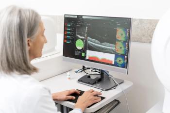
Dr. Clark reported findings from a study in which a commercially available SDOCT platform (Spectralis, Heidelberg Engineering) was used to determine changes in retinal curvature across the vertical meridian and to assess the outer nuclear layer (ONL) thickness as a surrogate measurement of photoreceptor density.
Advantages of SD-OCT
The results showed the SD-OCT-based technique generated curvature measurements that corresponded to those previously reported using refractive methods, as well as in terms of asymmetry in the retinal neuronal layer.
"Our experience indicates SD-OCT can rapidly measure changes in the optical path length across the retina, as well as how much neural potential there is to sense the signal. This technology, however, allows researchers to do more than just compare asymmetries in the inferior and superior retina, as we explored in this study," said Dr. Clark, research optometrist at The Borish Center for Ophthalmic Research, Indiana University School of Optometry, Bloomington.
"It also allows comparisons and weighting of the signals received in the vertical and horizontal meridians to investigate how they influence emmetropization, and it enables understanding of neural mass across the retina, which will be helpful for determining how far out in the periphery contact lenses or spectacles need to correct vision to prevent myopic progression. With the ability to measure changes in neural density, we can also monitor the safety of new technology lenses. Follow-up of potential changes in cone density will be important to make sure that the induced blur does not have a detrimental effect on neurons in the periphery."
Dr. Clark also acknowledged, however, that application of SD-OCT to this field of research is not without limitations.
"It is a paraxial measurement of the optical path length and does not take into account the optics of the full pupil, for which a wavefront sensor is needed, and our methods are labor-intensive and require extensive data analysis. We are validating an automated segmentation system that will make the process much less time-consuming," he explained.
Study methodology
SD-OCT images were acquired using 30 degree vertical scans through the center of the fovea. Central refraction and partial coherence interferometry measurements were also obtained since OCT does not give refractive error data. The researchers manually segmented the images to separate out the different retinal layers and focused on the RPE/photore-ceptor interface for determining curvature.
Their methods controlled for tilt, which can be artificially induced by OCT misalignment, along with magnification and decen-tration effects to ensure the measurements used the same foveal reference point. Samples were taken at seven locations at 5-degree steps from the foveal center, and the data in microns was converted to diopters to allow comparison with results from previously reported studies.
Compared with data published by Atchison et al., selected because it represents the largest dataset with vertical meridian data, there were no differences in the overall curvature measurements for the two study groups. Similarly, comparisons of curvature data for the superior and inferior fields were consistent with published information from Atchison et al. and others in showing that the superior retina is slightly more myopic than the inferior retina with the difference between the two locations being statistically significant.
Superior, inferior data
ONL thickness measurements also showed asymmetry, corresponding to previous reports, and with thickness being greater in-feriorly than superiorly.
"This may indicate higher neural potential of the inferior retina to receive the defo-cus signal. These two variables, however, are actually in opposition to each other because the refractive data show the superior retina has more defocus than the inferior retina," Dr. Clark said.
In an initial attempt to model blur signal strength, Dr. Clark and colleagues calculated a parameter they termed "neural blur strength" that was derived as the product of ONL thickness and defocus in the same location. The results showed there were no statistically significant differences in neural blur strength between the inferior and superior fields.
"This is a very simple attempt to model blur signal strength, and we recognize it should be refined with the addition of other factors, including off/off field receptors, blur spot size, and luminous levels. Our initial data, however, suggest that this may not be important because there is still a myopic defocus, or a relative stop signal in both meridians," he said.
FYI
Christopher C. Clark, ODE-mail:
Dr. Clark has no financial interest in the subject.
Newsletter
Want more insights like this? Subscribe to Optometry Times and get clinical pearls and practice tips delivered straight to your inbox.


