The role of lid hygiene in ocular surface disease
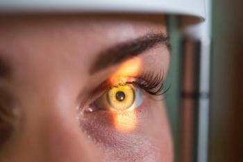
The ocular surface encompasses not only the cornea, but the all-important supporting conjunctiva that is divided into the bulbar, limbal, palpebral, forneaceal, and marginal zones.
Lid margin dysfunction is an important factor in the development of dry eye disease.1
The ocular surface encompasses not only the cornea, but the all-important supporting conjunctiva that is divided into the bulbar, limbal, palpebral, forneaceal, and marginal zones. The maintenance of the exquisite architecture of “the margin” is essential to proper tear and surface homeostasis.
Gentle but efficient lid hygiene promotes lid margin wellness.2
The complex lid margin
A brief review of lid margin anatomy and physiology is in order to better appreciate its elaborate complexity.
Let’s assume for this exercise that we have a healthy individual with an intact lid/blink system that effectively spreads the tear film across the ocular surface and efficiently pumps “spent” tears into the nasolacrimal system.
To do this, the lids must have normal tone with neither laxity nor notching and must coordinate with the blink to funnel tears toward the puncta for clearance. The puncta must be patent and large enough to accommodate the tear volume. Conjunctiva must neither billow or be redundant blocking the puncta nor interrupt the tear prism.
Of course, critical to this system are tears of normal composition and volume. When balanced, the tears support the ocular surface with nutrients, growth factors, antimicrobials, and a host of other molecules necessary for continued ocular surface health and function. When altered by disease , the tears can carry a battery of inflammatory agents that can ultimately impact the integrity of every unit of the ocular surface system from the eyelashes to the puncta.
Eyelashes grow in imperfect rows of five to six in the upper lid and three to four in the lower lid. There mean number is 90 to 160 in the upper lid and 75 to 80 in the lower lid; their length 8 to 12 mm in the upper lid, 6 to 8mm in the lower lid.
The skin of the eyelids is the thinnest of the body (< 1 mm). The nasal portion of the eyelid skin has finer hairs and more sebaceous glands than the temporal aspect, making this skin smoother and oilier.
Related:
Apocrine sweat glands of Moll are coiled glands in the dermis that empty via a ductile into the hair follicle. Sebaceous glands at the root of the hair follicle are holocrine glands that shed the entire epithelial cell along with secretory products of complex oils, fatty acids, wax, and cholesterol esters that is sebum. A large sebaceous gland is associated with each hair follicle and empties its secretions directly into the follicle. Additional small sebaceous glands of Zeis are present between lash follicles and dischreg their contents directly onto the skin surface. Its main purpose is to make the skin and hair waterproof and to protect them from drying out.
It is thought that overpopulation of Demodex mites precipitates pathological changes of the eyelids/eyelashes. These changes are consequences of blockage of the follicles and tubules of sebaceous glands by the mites and by reactive hyperkeratinization; epithelial hyperplasia from micro-abrasions caused by the mite’s claws; the mites acting as bacterial vectors; the host’s inflammatory reaction to the presence of parasite’s chitin as a foreign body; and stimulation of the host’s humoral responses and cell-mediated immunological reactions in response to the mites and their waste products.3 We can appreciate that maintaining the proper balance of these acarids is critical to overall normal lid/ocular surface function.4
Related:
Staphylococcal blepharitis is believed to be associated with staphylococcal bacteria on the ocular surface. However, the mechanism by which the bacteria cause symptoms of blepharitis is not fully understood.5
Early in our lives, bacteria from the environment colonize our conjunctiva, corneal surface, and associated tissues, including the eyelid and lacrimal systems. It is estimated that more than 200 species of bacteria commonly inhabit the human conjunctival mucosa.6
The ocular surface is chock-full of nutrients to sustain resident bacteria; in fact, in a balanced and intact ocular surface system, commensal bacterial species may protect the ocular surface from pathogenic infection.7
Mobile and free-floating bacteria are called the “planktonic” form. Interestingly, the life cycle of most bacteria is in sessile aggregates: microbes most often construct and live in a complex, film-like meshwork known as a biofilm.
Related:
A biofilm is a structural community of bacterial cells enclosed in a self-produced polymeric matrix that can adhere to inert or living surfaces.8 The biofilm environment provides physical protection to bacteria and also allows them to communicate with each other (quorum sensing), which may lead to an increase in virulence and propensity to cause infection.9
When bacteria change from being planktonic to biofilm-forming, they undergo changes in gene expression. There is speculation that such alterations create a more virulent strain of the bacteria or cause a conversion from a commensal species to a more harmful form.10
Related:
With the constant physical disruption of blinking, tear exchange, tear anti-microbial agents, and enzymes and mucins, bacteria generally face a robust ocular surface defense system that prevents generating a biofilm. However, when abiotic surfaces such as contact lenses, ocular prostheses, corneal sutures, and punctal plugs are introduced, biofilm formation upon them becomes a greater concern.
How lid hygiene helps
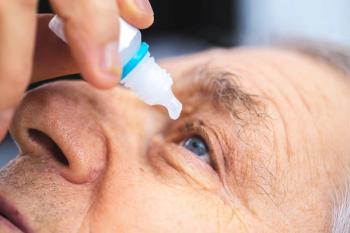
Appropriate lid hygiene practices are in order to lessen the bacterial load on the eyelid margin and eyelashes, aiding the natural ocular surface defense mechanisms. Prof. Benitez-Del-Castillo suggests that eyelid hygiene should be incorporated into a broader concept of eyelid health in which eyelid cleansing is part of a more complete program of care that includes screening and risk assessment, patient education, and coaching.2
Finally, remember that cosmetics have been documented to negatively impact the ocular adnexa, ocular surface, lacrimal system, and tear film.11
I recommend, and most patients prefer, commercially prepared lid hygiene products. Most of these are available as foaming cleansers or pre-moistened wipes, such as Ocusoft Lid Scrub Plus foam or Lid Scrub pads.
Additionally, in-office removal of eyelid/lash debris and de-bulking microbial load and biofilms can be achieved with Rysurg’s instrument BlephEx, which mechanically debrides treated surfaces.
Related:
There is much to learn about biofilm ecology and how it relates to bacterial virulence. In the meantime, it would seem prudent to clear away bacteria of concern before pathology sets in.
The benefits of eyelid hygiene/managing blepharitis are likely multifold. Reducing debris, allergens, and bacterial colonization on the eyelids reduces the risk for conjunctivitis and is thought to lower the chance of endophthalmitis after cataract surgery12 or from filtering blebs13 and can offer symptomatic relief of the deleterious effects of ocular surface disease and meibomian gland dysfunction.5
Indeed, it has been demonstrated that lid hygiene incorporated into therapeutic care for perennial conjunctivitis improves secondary dry eye symptoms and enhances the effectiveness of the treatment.14
Related:
Finally, remember that cosmetics have been documented to negatively impact the ocular adnexa, ocular surface, lacrimal system, and tear film.11
Protect the ocular health of your patients . Remind them of the importance of lid hygiene to promote eye wellness.
References
1. Knop E, Korb DR, Blackie CA, Knop N. The lid margin is an underestimated structure for preservation of ocular surface health and development of dry eye disease. Dev Ophthalmol. 2010;45:108-22.
2. Benitez-Del-Castillo JM. How to promote and preserve eyelid health. Clin Ophthalmol. 2012;6:1689-98.
3. Czepita D, Kuà ºna-Grygiel W, Czepita M, Grobelny A. Demodex folliculorum and Demodex brevis as a cause of chronic marginal blepharitis. Ann Acad Med Stetin. 2007;53(1):63-7; discussion 67.
4. Gao YY, Di Pascuale MA, Elizondo A, Tseng SC. Clinical treatment of ocular demodecosis by lid scrub with tea tree oil. Cornea. 2007 Feb;26(2):136-43.
5. Lindsley K, Matsumura S, Hatef E, Akpek EK. Interventions for chronic blepharitis. Cochrane Database Syst Rev. 2012 May 16;(5):CD005556.
6. Dong Q, Brulc JM, Iovieno A, Bates B, Garoutte A, Miller D, Revanna KV, Gao X, Antonopoulos DA, Slepak VZ, Shestopalov VI. Diversity of bacteria at healthy human conjunctiva. Invest Ophthalmol Vis Sci. 2011 Jul 20;52(8):5408-13.
7. Miller D, Iovieno A. The role of microbial flora on the ocular surface. Curr Opin Allergy Clin Immunol. 2009 Oct;9(5):466-70.
8. Costerton JW, Stewart PS, Greenberg EP. Bacterial biofilms: a common cause of persistent infections. Science. 1999 May 21;284(5418):1318-22.
9. De Kievit TR, Iglewski BH. Bacterial quorum sensing in pathogenic relationships. Infect Immun. 2000 Sep;68(9):4839-49.
10. Bjarsholt T. The role of bacterial biofilms in chronic infections. APMIS Suppl. 2013 May;(136):1-51.
11. Ng A, Evans K, North RV, Jones L, Purslow C. Impact of Eye Cosmetics on the Eye, Adnexa, and Ocular Surface. Eye Contact Lens. 2016 Jul;42(4):211-20.
12. Mamalis N, Kearsley L, Brinton E. Postoperative endophthalmitis. Curr Opin Ophthalmol. 2002 Feb;13(1):14-8.
13. Mac I, Soltau JB. Glaucoma-filtering bleb infections. Curr Opin Ophthalmol. 2003 Apr;14(2):91-4.
14. Ianchenko SV, Sakhnov SN, Malyshev AV, Fedotova NV, Orekhova OIu, Grishchenko IV. Treatment of chronic allergic blepharoconjunctivitis. Vestn Oftalmol. 2014 Sep-Oct;130(5):78, 80-4.
Newsletter
Want more insights like this? Subscribe to Optometry Times and get clinical pearls and practice tips delivered straight to your inbox.


