Study examines the effect of caffeine on retinal vessel density
A study1 that explored the effects of caffeine on systolic and diastolic blood pressure (SBP and DBP) and retinal vessel density (VD) assessed by optical coherence tomography angiography (OCTA) found that 200 mg of caffeine elevated the BP after 2 hours but did not impact the retinal VD, according to first author Mitchell Jacobs, MD, from the Department of Ophthalmology and Visual Sciences, University of Kentucky, Lexington, KY, and colleagues.
Previous studies, according to the investigators, have reported contradictory findings, with some suggesting adverse effects on the BP and cardiovascular risk, while meta-analyses proposed potential cardiovascular benefits.2-7 In contrast, the vascular and anti-inflammatory effects of caffeine are associated with a 65% decreased risk of dementia and Alzheimer’s disease.8,9
In the eye, caffeine can temporarily increase the intraocular pressure and reduce blood flow in critical ocular regions.10,11 Other studies using OCTA reported that caffeine consistently leads to decreased VD, suggesting a vasoconstrictive effect on the retinal circulation,12,13 according to Dr. Jacob and colleagues.
In light of these conflicting findings, the investigators used OCTA to determine how 200 mg of caffeine affects the SBP and DBP and VD in a prospective, randomized, double-blind placebo-controlled study. The study included 59 healthy coffee drinkers who ingested less than 136 mg of caffeine daily. Forty-two participants were randomly assigned into the caffeine group and received a 200-mg caffeine pill, and 17 were assigned to the placebo group.
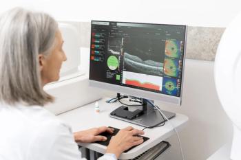
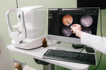
OCTA was performed at 60 and 120 minutes after the intervention. The VD was measured in the superficial capillary plexus (SCP) and deep capillary plexus (DCP) after the SBP and DBP measurements were recorded before each imaging session.
The findings showed that 2 hours after the intervention, the caffeine group had a significantly higher SBP (123 ± 7 mmHg) and DBP (81 ± 5 mmHg) compared to the control group (118 ± 7 mmHg, 77 ± 6 mmHg, respectively) (p =0.012, 0.023, respectively).
No significant differences were seen in the VD between the caffeine and placebo groups, regardless of whether the scans were centered on the macula or optic nerve head.
Dr. Jacobs and colleagues concluded that 200 mg of caffeine elevated the BP after 2 hours but did not impact the retinal VD. “This underscores the intricate relationship between caffeine, BP, and retinal vascular dynamics, prompting further exploration of their implications for ocular health, especially in subjects with vascular disease,” they commented.
References
Jacobs M, Demas N, Hemesath A, et al. Optical coherence tomography angiography: investigating vessel density changes induced by caffeine in healthy subjects. J Ophthalmol. 2024; published Oct 28;
https://doi.org/10.1155/2024/5597188 Zhou A, Hyppönen E. Long-term coffee consumption, caffeine metabolism genetics, and risk of cardiovascular disease: a prospective analysis of up to 347,077 individuals and 8368 cases. Am J Clin Nutr. 2023;109:509-516.
James JE. Chronic effects of habitual caffeine consumption on laboratory and ambulatory blood pressure levels. Eur J Cardiovasc Prevent Rehab. 1994;1:159–164;
https://doi.org/10.1177/174182679400100210 Vlachopoulos C, Hirata K, Stefanadis C, Toutouzas P, O’Rourke MF. Caffeine increases aortic stiffness in hypertensive patients. Am J Hypertens. 2003;16:63-66.
Wilson PWF, Bloom HL. Caffeine consumption and cardiovascular risks: little cause for concern. JAHA. 2016;5;
https://doi.org/10.1161/JAHA.115.003089 Caldeira D, Martins C, Alves LB, et al. Caffeine does not increase the risk of atrial fibrillation: a systematic review and meta-analysis of observational studies. 2013;99:1383-1389; doi: 10.1136/heartjnl-2013-303950
Miller PE, Zhao D, Frazier-Wood AC, et al. Associations of coffee, tea, and caffeine intake with coronary artery calcification and cardiovascular events. Am J Med. 2017;130:188–197.e5;
https://doi.org/10.1016/j.amjmed.2016.08.038 Vercambre MN, Berr C, Ritchie K, Kang JH. Caffeine and cognitive decline in elderly women at high vascular risk. JAD. 2013;35:413–421;
https://doi.org/10.3233/JAD-122371 ,Eskelinen MH, Kivipelto M. Caffeine as a protective factor in dementia and Alzheimer’s Disease. JAD. 2010;20:S167–S174;
https://doi.org/10.3233/JAD-2010-1404 Ajayi OB, Ukwade MT. Caffeine and intraocular pressure in a Nigerian population. J Glaucoma. 2001;10:25–31;
https://doi.org/10.1097/00061198-200102000-00006 Avisar RR, Avisar E, Weinberger D. Effect of coffee consumption on intraocular pressure. Ann Pharmacother. 2002;36:992–995.
https://doi.org/10.1345/1542-6270(2002)036%3A0992:eoccoi%3E2.0.co;2 Tugan BY, Subasi S, Pirhan D, et al. Evaluation of macular and peripapillary vascular parameter change in healthy subjects after caffeine intake using optical coherence tomography angiography. Indian J Ophthalmol. 2022;70:879-889;
https://pubmed.ncbi.nlm.nih.gov/35225536/ .Karti O, Zengin MO, Kerci SG, Ayhan Z, Kusbeci T. Acute effect of caffeine on macular microcirculation in healthy subjects: an optical coherence tomography angiography study. Retina. 2019;39:964-971; DOI: 10.1097/IAE.0000000000002058
Newsletter
Want more insights like this? Subscribe to Optometry Times and get clinical pearls and practice tips delivered straight to your inbox.


