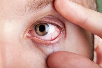
Think about the blink: Part 1
Blink rate and completeness matter most for comfort
All ODs are familiar with the Tear Film & Ocular Surface Society (TFOS) Dry Eye Workshop (DEWS) II report’s definition of dry eye disease as a “multifactorial disease of the ocular surface characterized by a loss of homeostasis of the tear film… in which tear film instability… plays an etiological role.”1
The blink plays a critical role in maintaining tear film stability and is crucial to sustaining tear film homeostasis. Yet, many practitioners fail to assess patients’ blink habits when examining eyes. As a dry eye practitioner, I have found that simply taking time to watch how a patient is blinking is a necessary step to correctly diagnosing and treating their ocular surface disease.
Functions of the blink
The blink accomplishes several tasks that are essential to preserving tear film homeostasis. During a complete blink, meibum is released from oil glands to the lid margin for dispersion onto the tear film. This is accomplished by the combined effort of the orbicularis oculi and the muscle of Riolan. The orbicularis compresses the tarsal plate to cause a milking effect on the meibomian glands, while the muscle of Riolan compresses the terminal ducts and acini of the glands. It is also believed that the muscle of Riolan keeps the meibomian glands closed to prevent leakage of meibum between blinks and during sleep.2,3
An essential function of the blink is that it causes the upper and lower tear film to mix. During a complete blink cycle, the upper lid thickens and remains perpendicular to the cornea, while the lower lid turns in slightly and thins out due to a slight nasal pulling toward the puncta. The result is that the upper lid does not actually touch the lower lid but overlaps it slightly. This creates a small space of approximately 0.7 mm that affects the combining of the upper and lower tear menisci, thereby causing the spread of meibum across the tear film and ocular surface. The upward movement of the lipid layer draws the aqueous tear film upward, as well.
The central lids do not actually touch during a complete blink, but the upper lid “overblinks” the lower one. Interestingly, the upper and lower tear films actually coalesce just before the blink is completed due to capillary attraction. Therefore, lipid spread actually occurs without the lid margins touching.4
The final essential function of the blink is to allow for tear drainage. As the upper lid moves down during a blink, the lower lid moves nasally, causing the thinning of the lower lid mentioned above. This nasal movement of the lower lid sweeps tear film debris toward the puncta for removal from the ocular surface. The upper and lower puncta make contact prior to the complete blink to drain the tears into the lacrimal sac. As long as the puncta meet, tear removal will occur even in the absence of a complete blink.5
Complications of incomplete blinks
Several complications may arise if a complete blink is not accomplished. When the upper lid does not meet and overlap the lower lid, less force is exerted on the glands, resulting in reduced meibum expression into the lower lipid reservoir. Additionally, there is a decrease in the replenishment of the lipid layer from the lower tear meniscus, resulting in uneven distribution of the lipid layer across the ocular surface. This leads to instability of the tear film and a lower tear break-up time. Additionally, chronic reduction in meibum expression leads to stagnation of meibum within the meibomian gland, resulting in gland obstruction and atrophy.2
It is important to note that not all blinks must be complete in order to maintain tear film homeostasis. A study by Korb et al found that 80 to 90 percent of blinks were complete in normal patients.5 Another study by Hirota et al observing blink patterns of computer users found that the tear film remained stable even with a reduced blink rate as long as most blinks were completed. However, a predominance of partial blinking resulted in an unstable tear film and a reduction in tear break up time.
Although the definition of partial blinking varies in literature, this study defined incomplete blinks as those in which the eyelid failed to cover the pupil.6
Take time to observe
It is important to take time to observe a patient’s blink pattern and see if the upper lid is actually making it to the lower lid. I have treated many patients, even children, with dry eye disease that is solely a result of incomplete blinking. And I have treated many more patients who exhibit incomplete blinking in combination with aqueous deficient or evaporative dry eye disease.
In order to fully restore tear film homeostasis, it is necessary to counsel patients on the importance of developing better blinking habits. This can be accomplished by training the patient to take regular breaks and initiate several forceful, sustained blinks, which will increase the lipid layer.
I love when I am able to show a patient that she is not blinking fully. Did you notice the lack of a complete blink in the video at the top? Here it is again in slow motion.
So, when you have a patient with complaints of ocular discomfort behind your slit lamp, make sure to pause for 15 to 20 seconds and assess his blink pattern.
In the upcoming installment, I will discuss causes and treatments of partial blinking. What you find may surprise you as well as your patients.
References
1. Craig JP, Nichols KK, Akpek EK, Caffery B, Dua HS, Joo CK, Liu Z, Nelson JD, Nichols JJ, Tsubota K, Stapleton F. TFOS DEWS II Definition and Classification Report. Ocul Surf. 2017 Jul;15(3):276-283.
2. Jie Y, Sella R, Feng J, Gomez ML, Afshari NA. Evaluation of incomplete blinking as a measurement of dry eye disease. Ocul Surf. 2019 Jul;17(3):440-446.
3. Knop E, Knop N, Millar T, Obata H, Sullivan DA. The international workshop on meibomian gland dysfunction: report of the subcommittee on anatomy, physiology, and pathophysiology of the meibomian gland. Invest Ophthalmol Vis Sci. 2011;52(4):1938-1978. Published 2011 Mar 30.
4. Pult H, Korb DR, Murph PJ, Riede-Pult BH, Blackie C.
5. Korb DR, Blackie CA, McNally EN. Evidence suggesting that the keratinized portions of the upper and lower lid margins do not make complete contact during deliberate blinking. Cornea. 2013 Apr;32(4):491-5.
6. Hirota M, Uozato H, Kawamorita T, Shibata Y, Yamamoto S. Effect of incomplete blinking on tear film stability. Optom Vis Sci. 2013 Jul;90(7):650-7.
Newsletter
Want more insights like this? Subscribe to Optometry Times and get clinical pearls and practice tips delivered straight to your inbox.


