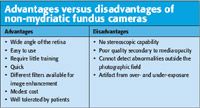
Why non-mydriatic cameras will not replace dilated fundus exams
The sensitivity and specificity of digital photography techniques - including non-mydriatic photography - are not as high as those of traditional fundus evaluation.
Baltimore-The sensitivity and specificity of digital photography techniques-including non-mydriatic photography-are not as high as those of traditional fundus evaluation. Nevertheless, these techniques may be useful in the future for screening and treatment trials, said Mahsa Salehi, OD, FAAO, here at the 5th annual Evidence Based Care in Optometry conference.

The AOA guidelines "mention that fundus photography provides documentation and is the best routine approach to establish a baseline for routine comparisons," she said. "They further point out that fundus photography is a more reproducible technique than the clinical exam for detecting posterior segment disease. It is not, however, medically necessary to document the existence of a condition, but medically necessary to establish the baseline to judge later if a disease is progressive." However, she noted that most third-party payers will not cover retinal photography alone without a concurrent eye examination.

Diabetic retinopathy (DR) continues to be one of the leading causes of blindness in the United States despite highly effective treatments. "We need to improve our ability to detect the onset of vision-threatening diabetic retinopathy by increasing our rates of screening of persons with diabetes," Dr. Salehi said, "especially for countries or areas that are not privileged and do not have as much access to ophthalmic care."
The gold standard for the documentation of DR consists of color-film stereoscopic photography of the seven standard fields, as the Early Treatment of Diabetic Retinopathy Study (ETDRS) established. "Photo screening, however, will not always detect the subtle signs and changes of DR, such as retinal thickening, but a success rate of 80% to 92% in detecting DR is claimed by researchers," Dr. Salehi noted.
Newsletter
Want more insights like this? Subscribe to Optometry Times and get clinical pearls and practice tips delivered straight to your inbox.