Consider the whole patient when treating glaucoma

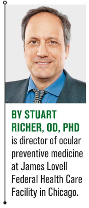
Stuart Richer, OD, PhD

ODs like to think that excellence in utilizing technology, prescribing pharmaceuticals, and making clinical decision(s) unites them in preventing human suffering and loss of vision. Nowhere is this more critical than in the diagnosis and treatment of glaucoma.
Yet, it may be shocking to hear that at a recent optometric continuing education meeting, one seasoned ophthalmologist told an audience that “based on multiple clinical studies, 20 to 59 percent of treated glaucoma patients lose visual field abiding by the standard of care.”1
With an increasing emphasis on technology over observation-and paraphrasing Mark Twain-are we looking under a light post for support rather than illumination?
Stewart Duke Elder, MD (1878-1978), a Scottish ophthalmologist, authored several textbooks, including System of Ophthalmology.2 Therein he states that glaucoma is a disease of the human body.
It appears the legendary “doctor’s doctor” had it right all along.
Previously by Dr. Richer: Intersection of artificial intelligence with epigenetics
Pulse pressure and predictive biomarkers
One place to start are seemingly simple blood pressure readings. The difference between systolic and diastolic blood pressure is known as “pulse pressure,” and it is associated with the degree of atherosclerosis.
While the pulse pressure typically widens with age, Westernized diets, and environmental stressors, readings wider than 40 mm Hg are an indication of compromised blood flow to end organ tissues such as the eye, particularly if endothelial function and autoregulation of blood flow is compromised. This is where preventative and environmental medicine come to the rescue.
The Health Studies Collegium lists eight predictive biomarkers that predict 92 percent of the epigenetic risk of all diseases, including cardiovascular disease.
These eight factors include:3
• Hemoglobin (Hb)A1c (dysglycemia)
• High-sensitivity C-reactive protein (hsCRP) (inflammation/repair deficit)
• Homocysteine (oxidant status, undermethylation)
• RBC omega-3 fatty acid index (chronic vascular disease and inflammation)
• 25 vitamin D liver reserve status (inflammation, atherosclerosis, survival)
• Early morning urine pH (acidity/alkalinity balance and magnesium/potassium mineral electrolyte status)
• Lymphocyte response antigens (immune tolerance)
• Antioxidant status
Unfortunately, most primary care providers measure only a few of these.
Related: Keep an eye on link between glaucoma and blood pressure
Magnesium status
Magnesium (Mg) is the fourth most abundant mineral in the body. Based upon Mg’s 300 enzymatic functions, it plays a seminal role in prevention and treatment of a plethora of diseases.
It is essential for the regulation of muscular contraction, blood pressure, insulin metabolism, cardiac excitability, vasomotor tone, nerve transmission, and neuromuscular conduction.4
Low levels of serum magnesium, manifested by overly acidic early morning urine pH, have been associated with a number of chronic diseases, such as Alzheimer's disease, insulin resistance/type 2 diabetes mellitus, hypertension, cardiovascular disease (such as heart attack and stroke), migraine headaches, and attention deficit hyperactivity disorder (ADHD).3
Mg has been shown to improve blood flow by modifying endothelial function and endothelial nitric oxide pathways. Mg also exhibits a neuroprotective role by blocking N-methyl-D-aspartate receptor-related calcium influx and inhibiting the release of glutamate, hence protecting cells against oxidative stress and apoptosis.5 Measuring the blood serum status of this oft-forgotten mineral costs $10.
Related: 6 healthy habits that don't cost a fortune Compromised sleep quality
In a previous department-“Sleep apnea: More than a snore and floppy eyelids,” (December 2018)-I discussed the association of ocular diseases like non-arteritic anterior ischemic optic neuropathy (NAION) and glaucoma with sleep apnea during a time of day when the supine body is already prone to low blood pressure and high intraocular pressure (IOP).
In the Maracaibo Aging Study, extreme decreases in night-time systolic and diastolic blood pressure (>20 percent compared with daytime blood pressure) were significant risk factors for glaucomatous damage, with dramatic odds ratios of 19.78 and 5.55, respectively.6
Another recent study suggests that sleep itself is important with a difference in melatonin production between high IOP glaucoma (low melatonin) and low-tension glaucoma (lower than normal, higher melatonin).7Related: Involve patients in monitoring IOP
Wrong lamp post
There is much we don’t know about glaucoma. Visual fields often worsen despite our fastidious efforts to lower IOP using drugs while repeating visual fields ad nauseum and monitoring the optic nerve head (and retinal ganglion cell layer) with the finest of imaging technologies.
Yet it is important to obtain adequate information about the latest scientific evidence found within peer-reviewed glaucoma publications to properly advise patients.8 But it is equally important to think across multiple medical disciplines beyond ophthalmology. Perhaps the real war should be against aging and combating multisensory impairment.9-11
If these observations are true, then our armamentarium could include more attention to the nuances of blood pressure measurements, proper sleep, exercise, stress reduction, and meeting universal predictive epigenetic health biomarkers. Perhaps low cerebrospinal fluid pressure is also involved.
If healthcare professionals, including primary-care ODs, fail to consider the whole patient, we will continue to meet clinical endpoints like IOP while still losing the war against glaucoma.
Read more nutrition articles here
References:
1. Teplick S. Presented at 2019 Island Eyes Conference. Pacific University College of Optometry. Jan. 25, 2019; Kohala Coast, Hawaii.
2. Lyle TK, Miller S, Ashton NH. Biographical memoirs of fellows of the Royal Society. William Stewart Duke Elder, 22 April 1898-27 March 1978. 26 Nov: 85-105, 1980.
3. Jaffe RM. Predictive biomarker tests: Use and application for evidence-based care. Available at: http://healthstudiescollegium.org/wp-content/uploads/2016/06/HSC_PredictiveBioMarkers_v211.pptx . Accessed 4/12/19.
4. Gröber U, Schmidt J, Kisters K, Magnesium in prevention and therapy. Nutrients. 2015 Sep 23;7(9):8199-226.
5. Ekici F, Korkmaz Å, Karaca EE, Sül S, Tufan HA, Aydin B, Dileköz. The role of magnesium in the pathogenesis and treatment of glaucoma. Int Sch Res Notices. 2014 Oct 13; 745439.
6. Melgarejo JD, Lee JH, Petitto M, Yépez JB, Murati FA Jin Z, Chavez CA, Pirela RV, Calmon GE, Lee W, Johnson MP, Mena LJ, AI-Aswad LA, Terwilliger JD, Allikmets R, Maestre GE, De Moraes CG. Glaucomatous optic neuropathy associated with nocturnal dip in blood pressure: findings from the Maracaibo Aging Study. Ophthalmology. 2018 Jun;125(6):807-814
7. Kim JY, Jeong AR, Chin HS, Kim NR. Melatonin levels in patients with primary open-angle glaucoma with high or low intraocular pressure. J Glaucoma. 2019 Feb;28(2):154-160.
8. Antón-López A, Moreno-Montanes J, Duch-Tuesta S, Corsino Fernandez-Vila P, Garcia-Feijoo J, Milla-Grino E, Munoz-Negrete FJ, Pablo-Julvez L, Rodriguez-Agirretxe I, Urcelay-Segura JL, Ussa-Herrera F, Villegas-Perez MP. Lifestyles guide and glaucoma (II). Diet, supplements, drugs, sleep, pregnancy, and systemic hypertension, Arch Soc Esp Oftalmol. 2018 Feb;93(2):76-86.
9. Costagliola C, Agnifili, L, Mastropasqua L, di Costanzo A. Low-tension glaucoma: An oxymoron in ophthalmology. Centers for Disease Control: preventing chronic disease. 2019;16:180534. doi: http://dx.doi.org/10.5888/pcd16.180534
10. Korsakova NV, The primary open-angle glaucoma: modern theory of development (literature review). Adv Gerontol. 2018;31(1):95-102.
11. Mancino R, Martucci A, Cesareo M, Giannini C, Corasaniti M,T Bagetta G, Nucci C, Glaucoma and Alzheimer’s disease: One age-related neurodegenerative disease of the brain. Curr Neuropharmacol. 2018;16(7):971-977.

Newsletter
Want more insights like this? Subscribe to Optometry Times and get clinical pearls and practice tips delivered straight to your inbox.