- March Digital Edition 2020
- Volume 12
- Issue 3
Pediatric headache: The vital role of the optometrist
Performing comprehensive examinations vigilantly is key
Optometrists play an important role in evaluating children who present with headaches. A comprehensive assessment is required to rule out neurologic signs, ocular pathology and binocular vision or accommodative dysfunction. Communicating findings to the patient’s medical doctor is also key.
A common reason that children are referred for an eye exam is a complaint of headaches. In fact, a survey found that 17 percent of 4- to 18-year-olds reported frequent, severe headaches and/or migraine in the previous year.1 Specifically, the prevalence of headaches was 4 percent of 4- to 5-year-olds and increased to 25 percent of 12- to 18-year-olds.
Often, pediatricians are tasked with determining if headaches are primary (tension, migraine) or secondary (organic, vascular, infectious, ocular, etc.). Before referring to pediatric neurology, rule out common causes of secondary headaches. This is where optometry comes in: ODs have a great opportunity to show colleagues their knowledge.
Related:
A comprehensive history is one of the most important components of headache evaluation. Discovering the temporal pattern of the headache is essential. These patterns can be divided into five categories in children: acute, acute-recurrent, chronic-progressive, chronic-nonprogressive, and mixed (Figure 1).2
Of these temporal patterns, chronic-progressive is the most ominous and should alert the examiner to be suspicious of organic causes (such as neoplasia, altered intracranial pressure, hemorrhage). Chronic-nonprogressive and mixed headache patterns are more likely to have ocular causes (such as uncorrected refractive error, binocular vision or accommodative dysfunctions).
Other factors to consider strongly as role players in pediatric headache are social stressors. These can include school, social media, friends, and drugs/alcohol. In addition, this can be compounded by lack of sleep or irregular sleep schedules, diet/nutrition, and dehydration. Be sure to include these components in the headache history.
Related:
The ocular exam
Just like any other examination, a headache evaluation begins with the basics: visual acuity, pupils, and extraocular motility testing. The following findings during assessments of these systems would be causes for alarm and would warrant further investigation:
Visual acuity that is reduced with no apparent refractive, amblyopic, or pathologic cause
Presence of anisocoria (non-physiologic) or an afferent pupillary defect
Restriction of movement on extraocular motility testing
Related:
A comprehensive evaluation of a headache patient should include an assessment of ocular health and visual field (automated if the patient is able). Visual field is an important tool to evaluate the integrity of the visual pathway. Neurologic visual field loss (such as homonymous hemianopsia, bitemporal hemianopsia) would indicate a likely organic cause of headache. Increased intraocular pressure (IOP) and anterior and/or posterior segment inflammation should be ruled out as a cause of pain that may be interpreted as headache by a child. The most ominous ocular finding to rule out is papilledema, which indicates increased intracranial pressure.
While it is important to rule out ocular signs of emergent headaches, in my experience the exam is much more likely to find that a secondary headache with ocular etiology is caused by something easily diagnosed and treated within an optometric office.
Foremost, refractive error must be evaluated. Myopia and astigmatism are typically easy to detect, but hyperopia can be a little trickier. In some children, even small amounts of hyperopia can cause headaches. This hyperopia may be latent and not be apparent until the patient is cyclopleged (1% cyclopentolate recommended). In a headache patient, prescribing even low amounts of hyperopia is warranted to rule out that their refractive error may be the headache trigger.
Related:
A thorough evaluation of the binocular vision and accommodative systems can be fruitful in a child with headaches. A full assessment of these systems with normal values is summarized in Table 1.3
Related:
First, assess ocular alignment. A significant phoria (with poor vergence ranges) can be the cause of headache. Note these common binocular conditions that are frequently associated with headaches:
Convergence insufficiency: Exophoria greater at near
Convergence excess: Esophoria greater at near
Divergence excess: Exophoria greater at distance
Ensure you are vigilant with your cover test. The presence of a strabismus should trigger the examiner to perform comitancy testing-evaluating the deviation in multiple positions of gaze. An incomitant strabismus indicates a muscle (mechanical) or nerve (neurologic) problem. A forced duction test can be used to differentiate between these two causes. The presence of a cranial nerve palsy can often be seen during extraocular motility testing, so watch carefully. Typically, a patient with a new-onset strabismus would complain of diplopia, but children often do not complain of this unless specifically asked.
Related:
The vergence system should be assessed to evaluate capacity and flexibility. Normal vergence ranges are noted Table 1. But, it is important to always be mindful of Sheard’s criterion as well-the compensating vergence range (BO for exo, BI for eso) should be twice the blur value (or break value if no blur is present). Vergence facility is commonly overlooked, but an inability to change from a convergence to a divergence posture poses difficulty and can be symptomatic. Inadequate fusional vergence, with or without significant phoria, can cause headaches.
Related:
The accommodative system evaluation should include assessment of magnitude, flexibility, and accuracy. This will help to diagnose common accommodative conditions associated with headache:
Accommodative insufficiency: Reduced accommodative amplitude, difficulty with minus lenses
Accommodative spasm: Over-accommodation; lead on monocular estimated method (MEM), difficulty with plus lenses
Accommodative infacility: Reduced accommodative facility with difficulty on plus and minus lenses
Accommodative fatigue: Inability to sustain accommodation; lag on MEM that increases with time, amplitude of accommodation that recedes with repetition
Related:
Optometric treatments
Refractive, binocular vision, and accommodative problems can be managed within optometric practice. Many of these conditions can be treated or initially managed with glasses alone. Do not forget the power of lenses.
Related:
Many accommodative disorders (accommodative insufficiency, fatigue, and sometimes even spasm) can be treated with glasses as well. Giving the child a bifocal in his glasses to decrease the accommodative demand at near can make a significant difference. Additionally, high accommodative convergence/accommodation (AC/A) conditions also respond to added lenses-convergence excess with extra plus at near and divergence excess with extra minus at distance.
Binocular vision and accommodative conditions are also successfully managed with vision therapy (VT). It is the gold-standard treatment for conditions like convergence insufficiency4 and is used effectively to remediate many binocular vision, accommodative, or oculomotor deficiencies.5 Many patients who have completed a VT program will report a decrease in their symptoms, including headache. VT can be used in conjunction with lenses to manage these patients to improve their symptoms. Lenses can be used as a temporary solution to relieve symptoms immediately, while vision therapy is a longer-term solution to many of these visual conditions.
Related:
When to image?
The American Academy of Neurology and the Child Neurology Society published recommendations regarding neuroimaging as part of evaluation of headache in children and adolescents in 2002.5
Its recommendations are as follows:
- On a routine basis, neuroimaging is not indicated in children with recurrent headaches and a normal neurologic exam
- Neuroimaging can be considered in children with an abnormal neurologic exam, the coexistence of seizures, or both
- Neuroimaging can be considered in children who have historical data to suggest recent onset of severe headaches, change in type of headache, or associated factors suggestive of neurologic dysfunction
Related:
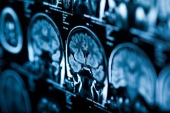
Another study looked at children who presented to the emergency department with the complaint of headaches and what signs or symptoms were most associated with them having a brain neoplasm versus clean neuroimaging.6 The findings show that the following were significant: neurologic signs (10.3x greater chance of neoplasm present), seizure (10.8x) and vomiting (6.6x). So, it is important to remember that neurologic signs and symptoms play a significant role in the decision to image a pediatric headache patient.
Although neurologists conduct a cursory evaluation of visual acuity, pupils, visual field, extraocular motilities, and gross funduscopic exam, this is optometrists’ specialty area. ODs are poised to evaluate the visual system and provide input on ocular neurologic signs that they may see. It is important to remember these neurologic findings should be communicated to the neurologist in order to facilitate neuroimaging. Or, if you are able, imaging can be ordered yourself due to:
- Acutely reduced visual acuity with no apparent cause
- New-onset anisocoria or the presence of an afferent pupillary defect
- Cranial nerve palsy
- Neurologic visual field loss
- Papilledema
- Non-ocular headache management
Related:
Sometimes it is not obvious whether the headache is due to the patient’s eyes. Perhaps the findings are borderline, and you are not convinced. In this case, a headache log can be useful to monitor headache characteristics such as frequency, duration, and what activities might trigger their onset.
Related:
For example, headaches that occur after reading are often visual, while headaches that wake a person from sleep are not. These logs are available as free apps (such as Migraine Buddy or iHeadache) that are easily accessible and make tracking seamless.
As healthcare providers, ODs can also counsel patients on lifestyle changes that can promote a healthier and more headache-free life. Suggest these SMART lifestyle changes:2
- Sleep: Get sufficient and appropriate sleep
- Meals: Ensure regular intake of healthy foods and good fluid intake
- Activity: Engage in regular and appropriate activity, neither excessive nor deficient
- Relaxation: Consider methods of stress management and relaxation
- Trigger avoidance: Recognize and avoid or manage situations that provoke headache
ODs are part of the team working toward getting to the root of the headache. Your findings will prove useful to the patient’s medical doctor. While it may help cross a differential off the list, it also could be the information the provider was waiting for to show medical necessity for neuroimaging.
Related:
References:
1. Lateef TM, Merikangas KR, He J, et al. Headache in a national sample of American children: prevalence and comorbidity. J Child Neurol. 2009 May;24(5):536-43.
2. Blume HK. Childhood headache: a brief review. Pediatr Ann. 2017 Apr 1;46(4):e155-e165.
3. Scheiman M, Wick B. Clinical Management of Binocular Vision: Heterophoric, Accommodative, and Eye Movement Disorders. Philadelphia: Lippincott Williams & Wilkins, 2013.
4. Convergence Insufficiency Treatment Trial Study Group. Randomized clinical trial of treatments for symptomatic convergence insufficiency in children. Arch Ophthalmol. 2008;126(10):1336-1349.
5. Scheiman M, Cotter S, Kulp MT, et al. Treatment of Accommodative Dysfunction in Children: Results from a Randomized Clinical Trial. Optom Vis Sci. 2011;88:1343–52.
6. Lewis DW, et al. Practice parameter: Evaluation of children and adolescents with recurrent headaches: Report of the Quality Standards Subcommittee of the American Academy of Neurology and the Practice Committee of the Child Neurology Society. Neurology. 2002 Aug 27;59(4):490-8.
7. Sheridan DC, Waites B, Lezak B, et al. Clinical factors associated with pediatric brain neoplasms versus primary headache: a case-control analysis. Pediatr Emer Care. 2017 Nov 14. doi: 10.1097/PEC.0000000000001347.
Articles in this issue
almost 6 years ago
New-onset, atypical retinopathy in a patient with diabetesalmost 6 years ago
Update on iris melanomaalmost 6 years ago
Use dark adaptation to screen before multifocal IOL implantationalmost 6 years ago
Unconventional clinical options for lowering IOPalmost 6 years ago
Optometry’s year can give ODs an edge with treatmentsalmost 6 years ago
New guidelines out for diabetes patient carealmost 6 years ago
The effect of contoured prism lenses on chronic headaches: a case studyalmost 6 years ago
Optometry’s role in multiple sclerosisalmost 6 years ago
5 tips to keep presbyopes in contact lensesalmost 6 years ago
How to create an ocular surface disease treatment protocolNewsletter
Want more insights like this? Subscribe to Optometry Times and get clinical pearls and practice tips delivered straight to your inbox.


