Blog: Recognize the signs of SND in your patients
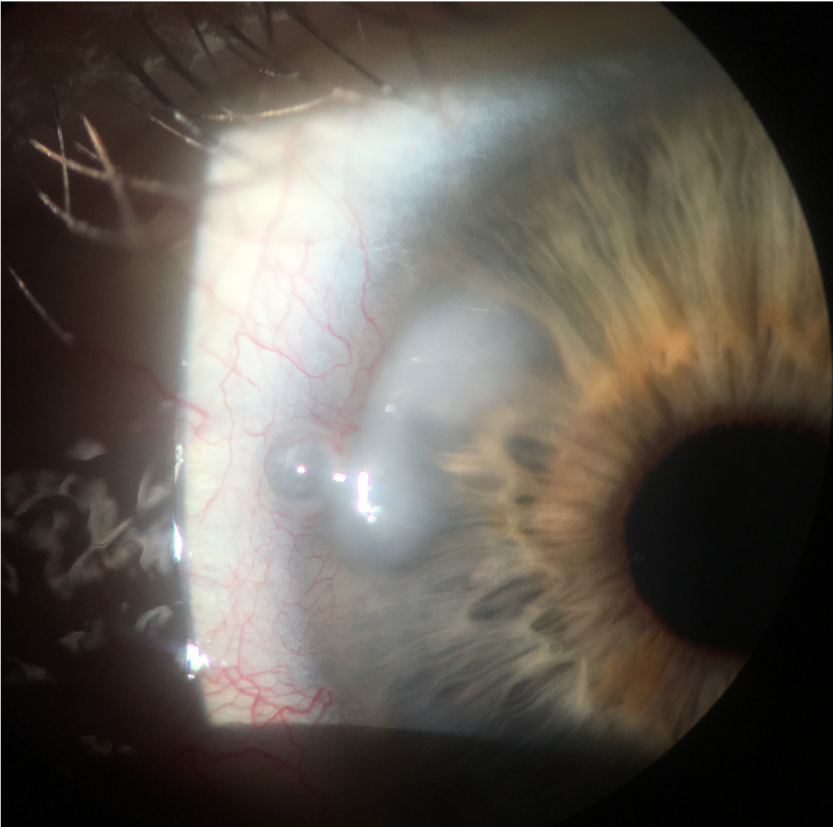
Salzmann's nodular degeneration can be identified by its blue-grey elevated nodular appearance of the cornea.


The views expressed here belong to the author. They do not necessarily represent the views of Optometry Times or Multimedia Healthcare.
As one of my mentors once said, “It’s not rare if it’s in your chair.”
Usually affecting Caucasian women in the fifth or sixth decade of life, Salzmann's nodular degeneration (SND) is a slowly progressive corneal condition that can be identified by its blue-grey elevated nodular appearance.
The disease affects females in 72 to 88 percent of reported cases, and often presents near the limbus or mid-peripheral cornea of males and females of all ages. About 63 percent of SND cases present bilaterally.1
Although this corneal degeneration can be sight threatening, recognizing the condition and identifying the underlying etiology is important. If identified and treated properly, SND has a positive prognosis.
Previously by Dr. Coats: GPC leads to contact instability
Case study
In one particular case, a 66-year-old Caucasian female presented with a gradually worsening foreign body sensation, epiphora, and light sensitivity. Compared to previous manifest refractions, she had acquired more irregular astigmatism, and her best corrected visual acuity (BCVA) had decreased from 20/25 to 20/40.
Upon slit lamp examination, a diagnosis of SND was made on the basis of the characteristic blue-grey bowtie appearance as well as the clinical history of progressively increasing irregular astigmatism associated with worsening of BCVA.
It is thought that Salzmann’s nodular formation is associated with inflammatory or non-inflammatory events involving trauma to the cornea, resulting in excess collagen or hyaline plaques replacing Bowman's layer.2
One hypothesis suggests that an enzymatic breakdown or destruction of Bowman’s membrane occurs with this initial corneal trauma, which can result in keratocytes from the posterior stroma migrating forward to create the superficial nodular areas.2
Microscopy of these nodules has the appearance of disorganized subepithelial collagen fibers and abnormally elongated basal epithelial cells.2
Common causes of chronic corneal trauma causing SND include meibomian gland dysfunction (MGD), keratoconjunctivitis sicca, chronic blepharitis, pterygium, exposure keratitis, trachoma, and interstitial keratitis.
Related: Why ODs should add meibography to their practices
Research
According to one report of 180 eyes identified with SND, chronic MGD was the most common comorbidity observed.1
Other causes of SND have been associated with epithelial basement membrane dystrophy (EBMD), soft monthly disposable contact lens wearers, and previous ocular surgery.1
Recent research findings have indicated an extremely high association between patients diagnosed with SND and osteoporosis, which suggests that SND may indicate an abnormality of the patient’s mucopolysaccharides.3
Anther case report suggests that SND may also be associated with chronic inflammatory diseases, such as Crohn’s disease.4
Management
Unfortunately, SND does not spontaneously resolve. It often ends up needing some level of treatment.
For asymptomatic patients, management can range from conservative observation to lipid-based lubrication (such as Systane Balance [Alcon], Refresh Optive [Allergan], and Tears Plus [Oasis Medical]).
Treatment options for symptomatic patients include a regimen of ointments, topical corticosteroids (loteprednol 0.5% [Lotemax, Bausch + Lomb]), and topical NSAIDs (bromfenac [Prolensa, Bausch + Lomb]) to relieve inflammation and pain.
For long-term treatment, topical dry eye therapies (cyclosporine 0.05% [Restasis, Allergan] and 0.09% [Cequa, Sun], lifitegrast 5% [Xiidra, Shire]) may be added to the regimen. These therapies have been shown to be effective in my clinic to help relieve symptoms for patients who are suffering from dry eye disease and/or MGD as the underlying culprit.
Related: Understanding and defining MGD
Depending on the underlying etiology, oral doxycycline may also be beneficial to reduce chronic inflammation.
Successful surgical options include superficial keratectomy (SK), with a subsequent phototherapeutic keratectomy (PTK) if irregular astigmatism is causing visual disturbances or if anterior haze persists. The recurrence rate of SND following PTK is low and estimated to be about 20 percent within 12 years of surgery.5
Other procedures-such as lamellar or penetrating keratoplasty-are reserved for nodules with deep stromal scarring. After removal of the nodules, amniotic membranes (Prokera [Bio-Tissue]) may help to restore corneal surface integrity by promoting healing and preventing haze.
Before a patient is referred for cataract surgery, superficial keratectomy of the astigmatism-inducing Salzmann’s nodules should always be performed.
Keratometry readings should be stable for four to six weeks in order to proceed with reliable intraocular lens (IOL) calculations. Premium and toric IOLs are usually avoided because of the risk of recurrent nodules and induction of postoperative astigmatism.
The patient mentioned earlier was able to undergo a successful superficial keratectomy procedure. Her vision has stabilized at 20/25 with a small myopic shift.
ODs may come across several cases of SND in their careers, so knowing what to look for and properly treating the underlying problem in a timely manner could influence a patient’s visual prognosis.
Disclosures: Dr. Coats is a member of Shire’s advisory board and speaks on behalf of the company.
References:
1. Farjo, A, Halperin G, Syed N, Sutphin JE, Wagoner MD. Salzmann’s nodular corneal degeneration clinical characteristics and surgical outcomes. Cornea. 2006 Jan;25(1): 11-15.
2. Stone D, Astley R, Shaver R, Chodosh J. Histopathology of salzmann nodular corneal degeneration. Cornea. 2008 Feb;27(2):148-51.
3. Karpecki PM, Schechtman DL. A look at Salzmann’s. Rev Optom. Available at: https://www.reviewofoptometry.com/article/a-look-at-salzmanns. Accessed 2/1/19.
4. Roszkowska AM, Spinella R, Aragona P. Recurrence of Salzmann nodular degeneration of the cornea in Crohn’s disease patient. Int Ophthalmolol. 2013 Apr;33(2):185-7.
5. Das S, Langenbucher A, Pogorelov P, Link B, Seitz B. Long-term outcome of excimer laser phototherapeutic keratectomy for treatment of Salzmann’s nodular degeneration. J Cataract Refract Surg. 2005 Jul;31(7):1386-91.
Newsletter
Want more insights like this? Subscribe to Optometry Times and get clinical pearls and practice tips delivered straight to your inbox.