The lowdown on hypoglycemia in patients with diabetes
Counsel your at-risk patients on strategies to minimize severe hypoglycemia.
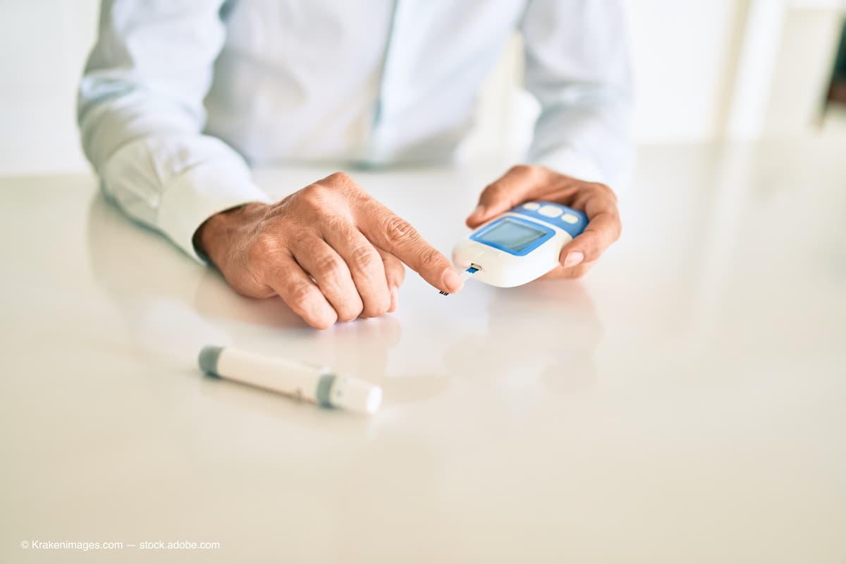
(Adobe Stock / Krakenimages.com)

When eye doctors think about metabolic disturbances caused by diabetes and the risk of eye disease, chronically elevated blood glucose levels that damage the retina often come to mind. Hyperglycemic insult to both vascular and neural elements of the retina has strongly been linked in multiple laboratory and clinical studies to the development and progression of diabetic retinal disease.1 But what about the antithesis of hyperglycemia—hypoglycemia? Does it play a role in the pathogenesis of diabetic retinopathy (DR)? Is low blood glucose something with which eye care providers should concern ourselves when we counsel our patients about risk mitigation? Let’s look at some of the evidence suggesting a definite affirmative response to these questions.
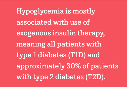
Hypoglycemia is mostly associated with use of exogenous insulin therapy, meaning all patients with type 1 diabetes (T1D) and approximately 30% of patients with type 2 diabetes (T2D). Severe hypoglycemia, defined as the presence of severe cognitive impairment requiring external assistance for recovery irrespective of the glucose level (typically < 54 mg/dL, which is what the American Diabetes Association defines as level 2 hypoglycemia), occurs in 3.3% to 13.5% of patients with T1D annually2 and 1% to 5% of patients with T2D annually in the US.3
Hypoglycemia is most common among older patients with multiple or advanced comorbidities, patients with long diabetes duration, or patients with a history of hypoglycemia. Insulin and sulfonylurea use, food insecurity, and fasting also increase hypoglycemia risk. Dead-in-bed syndrome refers to fatal cardiac arrhythmia due to severe hypoglycemia during sleep, typically with blood glucose levels below 30 mg/dL, and occurs primarily in younger children with T1D but also older adults with T1D or T2D due to neuroendocrine changes that reduce typical symptoms of hypoglycemia (hypoglycemia unawareness).4

Acute visual symptoms of hypoglycemia include blurred or dimmed vision, reduced contrast sensitivity, loss of visual field, diplopia, and even reduction in electroretinogram amplitude as a result of ATP (energy) depletion.5 In terms of DR, the Fremantle Diabetes Study conducted in Western Australia found that a history of hospitalization for severe hypoglycemia was the single biggest predictor of 2 or more lines of visual acuity loss in patients with diabetes for more than 4 years (5.6 times relative risk, whereas smoking tripled the risk and diabetic kidney disease nearly doubled the risk).6 Consistent with this finding, a large retrospective analysis of patients with T2D found that insulin use (the biggest risk factor for severe hypoglycemia) more than tripled the risk of developing proliferative DR (PDR) 5 years after diabetes diagnosis, whereas hemoglobin A1c of greater than 9% and renal disease more than doubled the risk.7
Recently, a study from Johns Hopkins University demonstrated a biochemical link between hypoglycemia and worsening DR.8 Retinal cells cultured in media with various levels of glucose (designed to simulate in vivo hypoglycemia) upregulated production of hypoxia-inducible factor 1a (HIF-1a) with subsequent increase in GLUT1 within the nuclei of Müller cells, resulting in downstream production of angiogenic proteins. This includes VEGF and ANGPTL4, both of which destabilize the blood-retinal barrier and promote abnormal neovascularization, which are the hallmarks of diabetic macular edema and PDR, respectively.
This phenomenon was more pronounced as levels of glucose in the culture medium decreased (ie, there was a dose-dependent response to increasing levels of hypoglycemia). Moreover, when Müller cells were placed under hypoxic stress—as happens in retinas exposed to prolonged hyperglycemia due to capillary closure—HIF-1a, VEGF, and ANG2 production significantly increased. Similarly, when rats were made hypoglycemic with administration of insulin, even for relatively short periods of time, Müller cells accumulated HIF-1a and angiogenic proteins massively increased.
The hypothesis here is that once DR develops due to long-standing diabetes and hyperglycemia, hypoxia from retinal capillary closure coupled with hypoglycemia results in the production of VEGF and other vasoproliferative mediators that worsen DR severity more than what would occur without hypoglycemic stress. From a practical standpoint, this means eye care providers should counsel patients with early nonproliferative DR to use strategies to minimize risk of hypoglycemia, including large glycemic fluctuations (cyclical hyperglycemia followed by hypoglycemia). For patients using insulin, continuous glucose monitoring (CGM) systems are especially useful for detecting and correcting hypoglycemia before it becomes severe, as all these devices provide an audible and/or vibratory alarm once low glucose thresholds are crossed. These devices help patients prevent severe hypoglycemia and can reduce mortality from dead-in-bed syndrome in high-risk patients.9
For patients using an insulin pump, use of an automated insulin delivery (AID) system that adjusts basal insulin delivery depending on current and predicted glucose levels within the hour is another way to avoid disabling hypoglycemia.10
(Author’s personal note: I have had type 1 diabetes for 54 years and have been wearing one of these systems [the Omnipod 5 AID pump combined with a Dexcom CGM] for about a month now and have experienced no hypoglycemia. Prior to use of the AID algorithm, I invariably experienced significantly low blood glucose levels during some nights, as well as in the afternoon while seeing patients).
For patients on noninsulin therapies capable of causing hypoglycemia by provoking insulin secretion from a functioning pancreas—primarily sulfonylureas (eg, glyburide, glipizide, 3glimepiride) and glinide nonsulfonylurea insulin secretagogues (eg, repaglinide and nateglinide)—most insurances will not cover CGM systems, but it may be worth the money for patients experiencing significant hypoglycemia to pay out of pocket for a device. At a minimum, patients experiencing hypoglycemia should be taught to perform home glucose spot monitoring with an inexpensive glucose meter and should work with their medical providers to access diabetes therapies that reduce risk of severe hypoglycemia (eg, ultra-fast acting insulins).

The American Optometric Association Clinical Practice Guidelines for Eye Care of the Patient With Diabetes Mellitus, 2nd edition, recommend that all optometrists keep a rapid-acting carbohydrate in-office for patients with diabetes who experience hypoglycemia during eye examination. With so many patients now using insulin, it’s a near certainty that each of us will encounter patients experiencing low blood glucose, and it is wise for every health care professional to be prepared for hypoglycemic emergencies. More broadly, we should ask high-risk patients about frequency and severity of hypoglycemia, counsel them on its prevention, and be even more vigilant for worsening diabetic retinal disease.
References
1. Shin ES, Sorenson CM, Sheibani N. Diabetes and retinal vascular dysfunction. J Ophthalmic Vis Res. 2014;9(3):362-373. doi:10.4103/2008-322X.143378
2. Pettus JH, Zhou FL, Shepherd L, et al. Incidences of severe hypoglycemia and diabetic ketoacidosis and prevalence of microvascular complications stratified by age and glycemic control in U.S. adult patients with type 1 diabetes: a real-world study. Diabetes Care. 2019;42(12):2220-2227. doi:10.2337/dc19-0830
3. Silbert R, Salcido-Montenegro A, Rodriguez-Gutierrez R, Katabi A, McCoy RG. Hypoglycemia among patients with type 2 diabetes: epidemiology, risk factors, and prevention strategies. Curr Diab Rep. 2018;18(8):53. doi:10.1007/s11892-018-1018-0
4. Parekh B. The mechanism of dead-in-bed syndrome and other sudden unexplained nocturnal deaths. Curr Diabetes Rev. 2009 Nov;5(4):210-5.
5. Khan MI, Barlow RB, Weinstock RS. Acute hypoglycemia decreases central retinal function in the human eye. Vision Res. 2011;51(14):1623-1626. doi:10.1016/j.visres.2011.05.003
6. Drinkwater JJ, Davis TME, Davis WA. Incidence and predictors of vision loss complicating type 2 diabetes: the Fremantle Diabetes Study Phase II. J Diabetes Complications. 2020;34(6):107560. doi:10.1016/j.jdiacomp.2020.107560
7. Gange WS, Lopez J, Xu BY, Lung K, Seabury SA, Toy BC. Incidence of proliferative diabetic retinopathy and other neovascular sequelae at 5 years following diagnosis of type 2 diabetes. Diabetes Care. 2021;44(11):2518-2526. doi:10.2337/dc21-0228
8. Guo C, Deshpande M, Niu Y, et al. HIF-1α accumulation in response to transient hypoglycemia may worsen diabetic eye disease. Cell Rep. 2023;42(1):111976. doi:10.1016/j.celrep.2022.111976
9. Hermanns N, Heinemann L, Freckmann G, Waldenmaier D, Ehrmann D. Impact of CGM on the management of hypoglycemia problems: overview and secondary analysis of the HypoDE Study. J Diabetes Sci Technol. 2019;13(4):636-644. doi:10.1177/1932296819831695
10. Giménez M, Conget I, Oliver N. Automated insulin delivery systems: today, tomorrow and user requirements. J Diabetes Sci Technol. 2021;15(6):1252-1257. doi:10.1177/19322968211029937

Newsletter
Want more insights like this? Subscribe to Optometry Times and get clinical pearls and practice tips delivered straight to your inbox.