- Vol. 11 No. 4
- Volume 11
- Issue 4
Case: New protocol for macular hole treatment
A 55-year-old female had been followed for several months for a macular hole in the right eye. She returned for a scheduled visit and reported no change in visual acuity- the left eye had been and remained uninvolved.
The patient’s medical history was significant for diabetes of an undetermined duration for which she was under the care of another physician. Medication for this condition, as well as for her diagnosis of systemic hypertension, was not revealed.
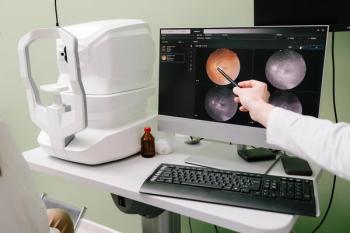
Visual acuity at the scheduled visit was 20/200 (20/80 EV) OD, and 20/25 OS. Fundus image of the right eye is seen in Figure 1.
Clinical assessment
On clinical evaluation, it was evident that full-thickness macular hole was present even absent the previous diagnosis and visual acuity.
Despite a duration of the diagnosis, however, there was no clinical evidence of a fluid surround-meaning that the full-thickness hole was limited to what was visualized.
Optic cube obtained from
Note that while clinical evaluation may allow observation of the hyaloid of the vitreous, the relationship between the vitreous and retina is emphasized in the optic-cube data.
This is a reminder of the presence of the mass of vitreous in relation to its remaining focal attachment at the macular surface.
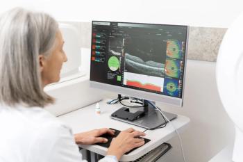
When interpreting optical coherence data, it may be tempting to analyze cross-sectional scans alone. While this can be useful, it could require interpolation to determine actual horizontal and vertical extent of a lesion.
The optic cube allows an overview that-when combined with the cross-sectional data-gives a more complete perspective.
Of further note is the remaining attachment between the detached vitreous and paramacular area of the right eye. This OCT finding corresponds with the white dot indicated in the fundus photograph. The macular hole is full in thickness and lacks a fluid surround. This correlation can be seen comparing the fundus image and the cross-sectional OCT (Figure 3).
The patient was seen in consultation with a retina specialist at this visit and offered a surgical option, which she accepted. The procedure consisted of parsplana vitrectomy with the placement of a gas bubble.
With just 48 hours of face-down positioning instead of the usual two-week interval, the patient tolerated the procedure well.
This new type of protocol is being tried by some retinal surgeons. The patient in this case is one of the first to use the protocol. It has proven to be useful for minimizing post-operative complications as well as producing positive surgical outcomes.
Postop
At the first postoperative visit two weeks later, visual acuity had not improved. However, anatomical closure of the macular hole had been achieved (see Figure 4). At a follow-up visit three months post-surgery, visual acuity improved to 20/40.
Case analysis
Two significant items are present in this case. First is the outside limit-fewer than 12 months-being favorable for surgical success due to the presence of the macular hole. While this has been reported for bilateral presentations, the results can be extrapolated to the monocular situation.1
Second is that on careful observation of the postoperative OCT, the external limiting membrane had been re-established.2 See Figure 4.
This is significant for a good prognosis of visual acuity improvement that occurred in the case. As is consistent with the improved visual acuity, the photoreceptor layer repopulated.
Interestingly, the fellow eye showed the presence of vitreomacular adherence (vitreomacular attachment without disruption of retinal anatomy).3 Vitreomacular traction (adherence between vitreous and retina with anatomical tissue disruption) places the patient at a 30 percent risk of macular hole formation in the following five years.4
Vitreomacular adherence carries a much lower risk of macular hole formation.4,5
Disclosures:
1. Chang E, Garg P, Capone A Jr. Outcomes and predictive factors in bilateral macular holes. Ophthalmology. 2013 Sep;120(9):1814-9.
2. Maheshwary AS, Oster SF, Yuson RM, Cheng L, Mojana F, Freeman WR. The association between percent disruption of the photoreceptor IS–OS and visual acuity in diabetic macular edema. Am J Ophthalmol. 2010;150:63–67.
3. Rodman JA, Shechtman D, Sutton BM, Pizzimenti JJ, Bittner AK; VAST Study Group. Prevalence of of vitreomacular adhesion n patients without maculopathy older than 40 years. Retina. 2018 Oct;38(10):2056-2063.
4. Philippakis E, Astroz P, Tadayoni R, Gaudric A. Incidence of macular holes in the fellow eye without vitreomacular detachment at baseline. Ophthalmologica. 2018;240(3):135-142.
5. Errera MH, Liyanage SE, Petrou P, Keane PA, Moya R, Ezra E, Charteris DG, Wickham L. A study of the natural history of vitreomacular traction syndrome by OCT. Ophthalmology. 2018 May;125(5):701-707.
Articles in this issue
almost 7 years ago
4 ways to use eye tracking in your practicealmost 7 years ago
How to see 50 patients a day at your practicealmost 7 years ago
Best practices for comanaging crosslinking patientsalmost 7 years ago
Cataract surgery comanagement from the other sidealmost 7 years ago
How to educate patients on risks of eyelash enhancementsalmost 7 years ago
Pros and cons of private equity firms in practice transitionsalmost 7 years ago
Know the connection between vitamin D and dry eye diseasealmost 7 years ago
Interest growing in virtual CEalmost 7 years ago
5 marketing tips to grow a practice without breaking the bankNewsletter
Want more insights like this? Subscribe to Optometry Times and get clinical pearls and practice tips delivered straight to your inbox.


