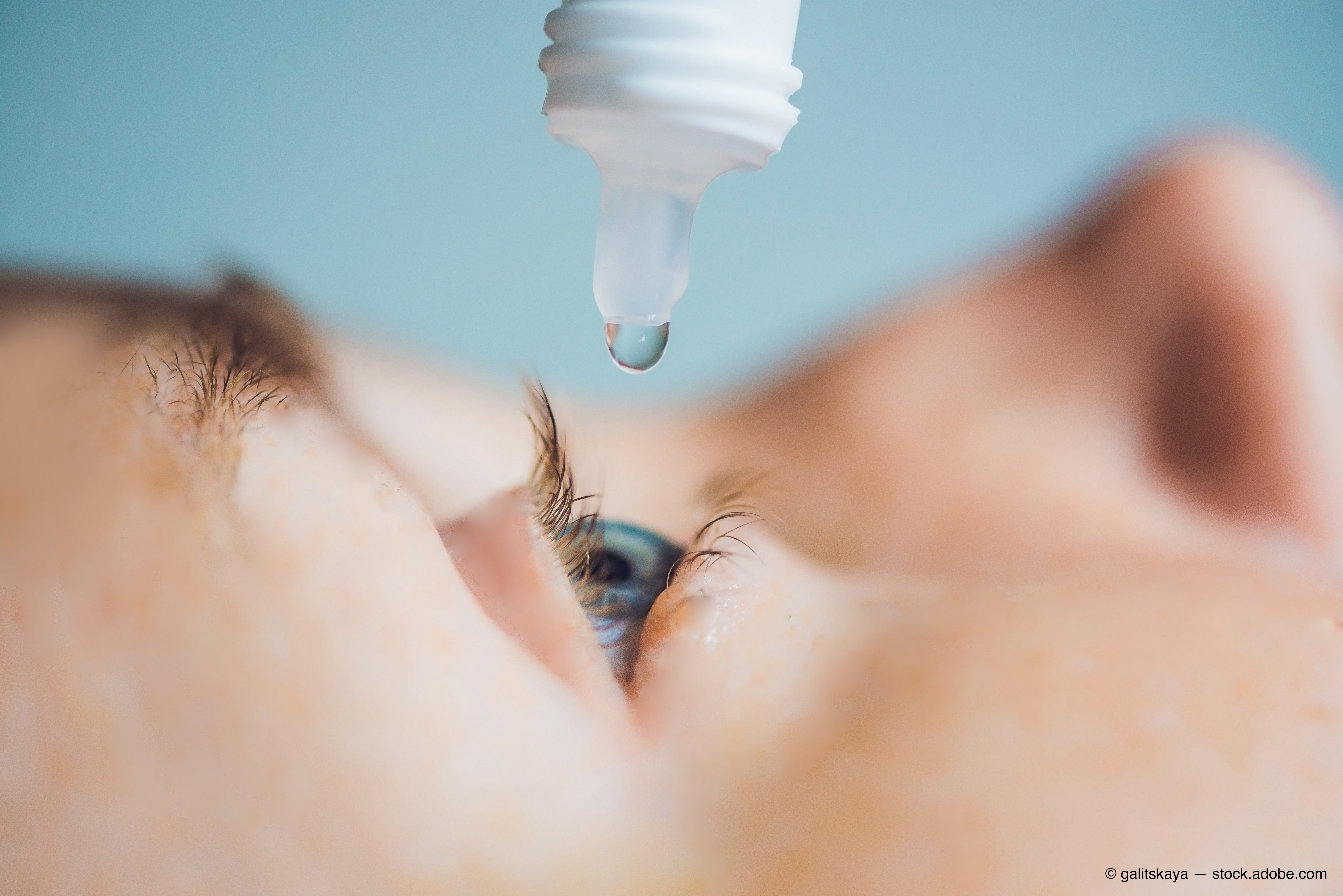
Know the connection between vitamin D and dry eye disease


I am fortunate to practice in a setting where my patient’s full medical and drug therapy is readily available. I have noticed that many patients (including myself) have been prescribed oral vitamin D supplements (cholecalciferol) to boost serum levels of vitamin D.
Vitamin D deficiency (VDD) and insufficiency (VDI) are increasing on a global level. VDD is associated with an increased risk of common chronic diseases such as bone metabolic disorders, tumors, cardiovascular diseases, and diabetes.1
Related: Sunlight and its effect on eye health
Study finds
A retrospective study demonstrated the increasing prevalence of VDD (28.9 percent) and VDI (41.4 percent) from 2001 to 2010.
Study participants who were black, less educated, poor, obese, current smokers, physically inactive, and infrequent milk consumers had a higher prevalence of VDD.2
In the dry eye space, a December 2018 study in Cornea investigated the efficacy of topical carbomer-based lipid-containing artificial tears (CLAT) and hyaluronate (HU) in patients with dry eye disease (DED) based on serum 25-hydroxyvitamin D (25HD) levels and cholecalciferol supplementation.
The data of this study suggests that the effect of topical CLAT and HU was dependent on serum 25HD levels. The authors conclude that cholecalciferol supplementation enhanced the efficacy of topical treatment and may be a useful adjuvant therapy for patients with DED refractory to topical lubricants.3
Epidemiological studies have linked VDD with other ocular diseases, such as myopia, age-related macular degeneration (AMD), diabetic retinopathy, glaucoma, retinoblastoma, and uveitis. However, the underlying mechanism is not yet fully understood.
Previously by Dr. Mastrota: How to recognize and manage digital eye strain
Case report
Parts of the human eye-including the epithelium of the cornea, lens, ciliary body, and retinal pigment epithelium, as well as the corneal endothelium, ganglion cell layer, and retinal photoreceptors-contain vitamin D receptors (VDR). Intramuscular injections of vitamin D were demonstrated to improve tear hyperosmolarity.4
The recently discovered anti-angiogenic, anti-inflammatory, and anti-neoplastic properties of calcitriol (the hormonally active metabolite of vitamin D) have shed some light on vitamin D’s role in the pathogenesis of ocular diseases.5
Interestingly, there has been an isolated, VDD-related case report of a 40-year-old patient with persistent bilateral ocular pain and discomfort for two years, for whom conventional management of dry eye had failed.
Related: Blog: Don't leave dry eye patients high and dry
Detailed ocular examination, meibography, and tear film evaluation were suggestive of bilateral meibomian gland dysfunction (MGD) and evaporative dry eye. Topical medication failed to alleviate the patient's symptoms.
Conventional management having failed, LipiFlow (Johnson & Johnson Vision) was performed, and topical therapy with cyclosporine 0.05%, steroids, and lubricating eye drops was initiated with incomplete symptomatic relief.
To identify the cause of pain, imaging was performed with in vivo confocal microscopy and anterior segment spectral domain optical coherence tomography (OCT). Systemic evaluation revealed severe VDD.
With parenteral therapy for VDD, there was a dramatic improvement in this patient's symptoms. The authors of this case study suggested that inflammation aggravated by VDD resulted in an altered epithelial profile, Bowman’s layer damage, recruitment of dendritic cells, and an altered subbasal nerve plexus features in this patient with chronic DED.6Related: How to address the three aspects of dry eye disease
Future for dry eye disease
Should vitamin D serum testing be part of ODs’ DED work-up? Is vitamin D supplementation prophylactic for DED? Conflicting data exists to support this notion.7,8
Awareness of the high prevalence of VDD and VDI among U.S. adults and related predictors could inform behavioral and dietary strategies for preventing VDD, monitoring VDI, and perhaps modulate its impact on/contribution to DED.
Eyecare practitioners hope that with more data they can answer these questions and have yet another avenue of therapy for ocular surface disease patients.
References:
1. Wang H, Chen Q, Li D, Yin X, Zhang X, Olsen N, Zheng SG. Vitamin D and chronic diseases. Aging Dis. 2017 May; 8(3): 346-353.
2. Liu X, Baylin A, Levy PD. Vitamin D deficiency and insufficiency among US adults: prevalence, predictors and clinical implications. Br J Nutr. 2018 Apr;119(8):928-936.
3. Hwang JS, Lee YP, Shin YJ. Vitamin D enhances the efficacy of topical artificial tears in patients with dry eye disease. Cornea. 2019 Mar;38(3):304-310.
4. Kizilgul M, Kan S, Ozcelik O, Beyesl S, Apaydin M, Ucan B, Cakal E. Vitamin D replacement improves tear osmolarity in patients with vitamin D deficiency. Semin Ophthalmol. 2018;33(5):589-594.
5. Skowron K, Pawlicka I, Gil K. The role of vitamin D in the pathogenesis of ocular diseases.
Folia Med Cracov. 2018;58(2):103-118.
6. Shetty R, Deshpande K, Deshmukh R, Jayadev C, Shroff R. Bowman break and subbasal nerve plexus changes in a patient with dry eye presenting with chronic ocular pain and Vitamin D deficiency. Cornea. 2016 May;35(5):688-91.
7. Yang CH, Albietz J, Harkin DG, KimLin MG, Schmid KL. Impact of oral vitamin D supplementation on the ocular surface in people with dry eye and/or low serum vitamin D.Cont Lens Anterior Eye. 2018 Feb;41(1):69-76.
8. Jeon DH, Yeom H, Yang J, Yang J, Song JS, Lee HK, Kim HC. Are serum Vitamin D levels associated with dry eye disease? Results from the study group for environmental eye disease. J Prev Med Public Health. 2017 Nov;50(6):369-376.

Newsletter
Want more insights like this? Subscribe to Optometry Times and get clinical pearls and practice tips delivered straight to your inbox.