The case of the scarred retina
Undiagnosed ocular trauma could lead to more complicated ocular problems for patients if left untreated. Leo P. Semes, OD, FAAO, looks at one case in which optical coherence tomography angiography (OCT-A) helped diagnose and treat a patient successfully.


My former ocular embryology teacher, Gilda Crozier, OD, FAAO, reminded her class during every lecture that “phylogeny replicates ontogeny” is similar to how newer technologies replicate clinical findings. To highlight this, I present an example of a patient who suffered blunt ocular trauma not involving the macula several decades prior to his exam.
Milky vision
This case highlights a 62-year-old male. At the time of the incident, he recalled his vision becoming milky, but it cleared within a week. He said his central vision was not affected permanently.
The patient described being evaluated by a general practitioner who did not create a management plan. He then followed up some months later with a general ophthalmologist who diagnosed him with a “scarred retina.”
A stable absolute visual-field defect corresponding roughly to the area of insult had been documented.
Exam shows drusen present
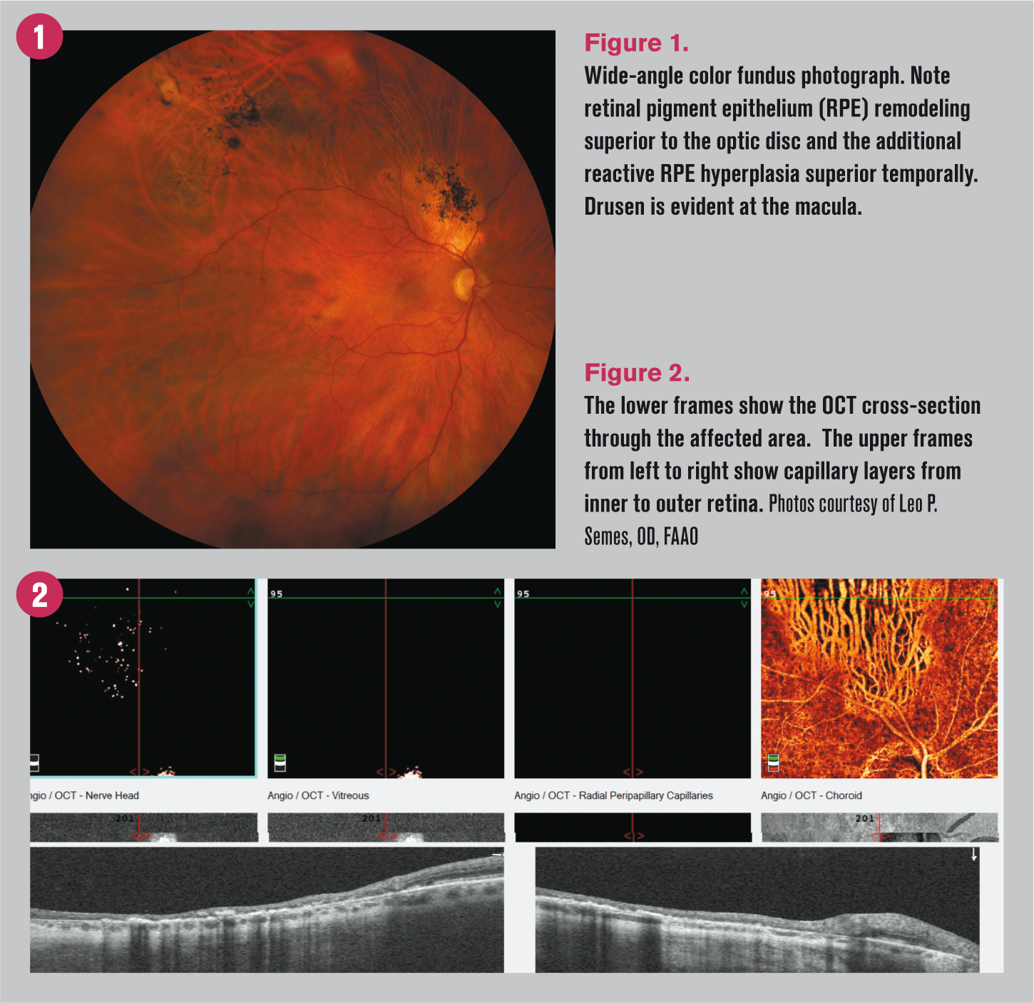
A decade following the trauma, he was observed to have a superior-temporal retinal hole that was prophylaxed with cryotherapy. The result of the blunt trauma was obvious above the optic disc and visible when observing the wide-angle photo.
The repaired retinal hole was captured in the superior temporal region (Figure 1).
In the intervening years, the patient had been a successful contact lens wearer and subsequently underwent successful cataract removal with intraocular lens (IOL) implantation nine years ago.
Visual acuity in this eye was 20/20 with -2.25 D refractive correction, which serves as his near eye in a monovision paradigm. The patient also presented with visible age-appropriate drusen in the macula.
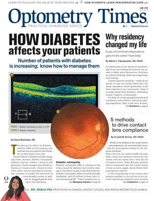
At the present evaluation, optical coherence tomography angiography (OCT-A) was performed (Figure 2). The OCT portion of the scan showed a thinned retina in the region of the retinal remodeling and peri-macular drusen consistent with the clinical observances.
The angiography component of the scan showed that the inner retinal layers were absent, and the layers through the inner retina failed to demonstrate evidence of capillary presence.
OCT-A helps with diagnosis
The significance of OCT-A is that it outlines capillary investment, presence, networks throughout the retina and choroid by means of motion perception of moving corpuscles.
OCT-A is a distinct technology from fluorescein angiography (FA). Fluorescein angiography looks at the integrity of the circulatory systems of the retina and choroid. OCT-A may make the greatest impact on clinical practice in early progressive situations of capillary loss.
With further research, we may be able to correlate reduced retinal blood flow as sentinels of neurodegenerative diseases.

Newsletter
Want more insights like this? Subscribe to Optometry Times and get clinical pearls and practice tips delivered straight to your inbox.