Contrast sensitivity manifests in glaucoma patient with no cataracts
A comment from a patient prompts a doctor’s change in testing and discussion.

Contrast sensitivity evaluates how well a person can distinguish an object from its background and it is an often overlooked symptom of glaucoma.
I have spoken to glaucoma patients and suspected glaucoma patients about the risk of visual field loss many times for several reasons. They need to know, they have a right to know, it is part of my fiduciary obligation, and it may improve compliance. I usually end the conversation by saying “if glaucoma goes untreated for long enough, it can affect your central vision, as well”.
A recent conversation with a glaucoma patient of mine led me to the unfortunate conclusion that I really need to rephrase my concluding remarks in this conversation (which I have often).
Related: Know what common glaucoma mistakes to avoid
Initial presentation
A Caucasian male who is now 51 years old first presented to me in 2015 for a comprehensive eye examination. He was a low myope and an early presbyope looking to update his current glasses. Best-corrected visual acuity (BCVA) was 20/20 OU. His family history was noncontributory.
Of note, his intraocular pressures (IOP) were 20 mm Hg OD and 21 mm Hg OS in the early afternoon. Upon posterior segment examination through dilated pupils, his right optic nerve head appeared suspicious for glaucoma with a slightly larger cup than his left optic nerve head. I thought his retinal nerve fiber layer, as examined with the use of a pre-corneal lens and a red-free filter, appeared asymmetric as well. There was no frank history of trauma. I took photos of his optic nerves and invited him back in a week or two for baseline glaucoma testing.
Related: Pediatric glaucoma: types, tests and treatments
Follow up
He eventually returned in October 2019. He apologized and stated he remembered our conversation about the need for glaucoma testing well but that he had just gotten busy and put it off until the present time. At that visit, his BCVA was 20/20 in each eye. IOPs were measured at 32 mm Hg OD and 16 mm Hg OS in the mid-morning.
Unfortunately, his right optic nerve appeared to be obviously glaucomatous with a vertical cup-to-disc ratio of 0.8 and a clearly evident notch superiorly at 11 a.m. His left optic nerve looked unchanged from his previous exam in 2015. Gonioscopy showed relatively symmetric angles open to the ciliary body OU with mid pigment. There was no evidence of angle recession. Central corneal thickness values were 533 μm OD and 537 μm OS.
Spectral-domain optical coherence tomography (OCT) studies OU showed a thin ganglion cell complex and diffuse retinal nerve fiber layer thinning OD. The OS study was essentially clear. Visual field studies were conducted, and the OD showed dense arcuate defects superiorly and inferiorly. OS was unremarkable.
Related: Misdiagnosis when clinical findings don’t make sense
I told the patient that he had unilateral open-angle glaucoma OD and that he needed to be treated. He consented to treatment, and I started him on a prostaglandin analog at bedtime OD with a target of 50 percent pressure reduction in that eye.
When he returned in a month IOPs were 14 mm Hg OD and 17 mm Hg OS at the same time of day. The dense arcuate defects in the OD visual field were repeatable, and the OS field remained unremarkable.
I congratulated him on his right eye’s remarkable response to glaucoma monotherapy and invited him to return again in 3 months for an IOP check. At that visit in February 2020, his IOPs were unchanged. It was at that visit that he intrigued me with a comment and a piece of paper he brought with him.
Related: 10 new treatments in eye care
Contrast sensitivity
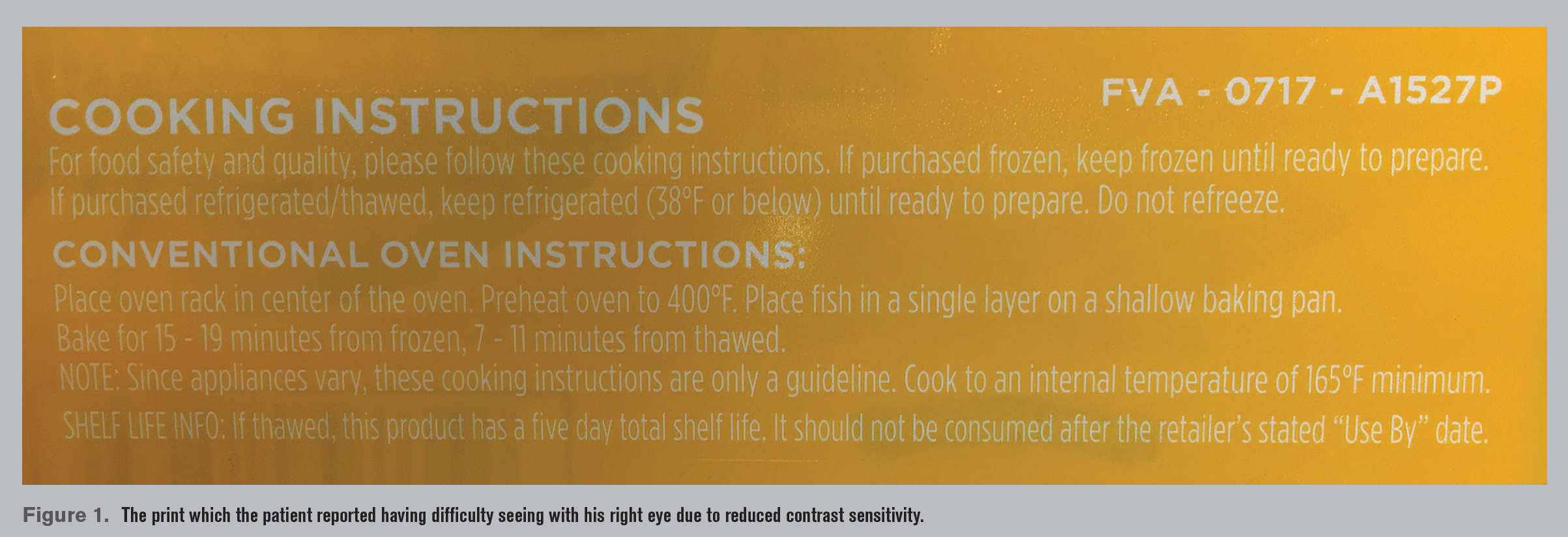
This man is a highly intelligent and astute observer He is an engineer with a PhD. He told me that he didn’t notice his visual field defect at all but that he did have a noticeable and bothersome change to his contrast sensitivity OD.
In fact, he brought in what he considered to be the quintessential demonstration of his complaint, an example of white writing against a yellow background (Figure 1).
He was kind enough to leave this with me so I could test contrast sensitivity on glaucoma patients. I thanked him and told him I would brush up on the topic. He said he will continue to self-monitor the disparity of contrast sensitivity between OD and OS and let me know if there is improvement in the future.
A pertinent negative finding to note is that this patient does not have a cataract in either eye (a potential lurking variable in patients for whom contrast sensitivity is a concern).
His BCVA is normal and symmetrical, and his cornea, aqueous, and vitreous humor are unremarkable.
The effect of glaucoma on contrast sensitivity is well documented.1-4 This parameter of visual function is related to numerous activities of daily living.5 Contrast sensitivity has been postulated to be a likely culprit of visual complaints in glaucoma patients with good visual acuity.6 Indeed, contrast sensitivity is an often-overlooked aspect of one’s visual quality of life.
I often talk about contrast sensitivity with patients with cataracts, but I am going to do a better job of addressing this aspect of glaucoma, as well. I am equipped to test for it, and I am going to do so more often after having this conversation with my patient. I am also going to rework how I explain visual function in glaucoma to include something about contrast sensitivity.
References:
1. Richman J, Lorenzana LL, Lankaranian D, Dugar J, Mayer J, Wizov SS, Spaeth GL. Importance of visual acuity and contrast sensitivity in patients with glaucoma. Arch Ophthalmol. 2010 Dec;128(12):1576-1582.
2. Ross JE, Bron AJ, Clarke DD. Contrast sensitivity and visual disability in chronic simple glaucoma. Br J Ophthalmol. 1984 Nov;68(11):821-827.
3. Atkin A, Bodis-Wollner I, Wolkstein M, Moss A, Podos SM. Abnormalities of central contrast sensitivity in glaucoma. Am J Ophthalmol. 1979 Aug;88(2):205-211.
4. Sample PA, Juang PS, Weinreb RN. Isolating the effects of primary open-angle glaucoma on the contrast sensitivity function. Am J Ophthalmol. 1991 Sep;112(3):308-316.
5. Liu J, McAnany JJ, Wilensky JT, Aref AA, Vajaranant TS. M&S Smart System contrast sensitivity measurements compared to standard visual function measurements in primary open angle glaucoma patients. J Glaucoma. 2017 Jun;26(6):528-533.
6. Hawkins AS, Szlyk JP, Ardickas Z, Alexander KR, Wilensky JT. Comparison of contrast sensitivity, visual acuity, and Humphrey visual field testing in patients with glaucoma. J Glaucoma. 2003 Apr;12(2):134-8.

Newsletter
Want more insights like this? Subscribe to Optometry Times and get clinical pearls and practice tips delivered straight to your inbox.