- October digital edition 2022
- Volume 14
- Issue 10
Current glaucoma treatments bring challenges
Optometrists must identify and mitigate complications for this patient base.
As optometrists, we are taught to treat and manage many ocular diseases. But 1 disease sits on a pedestal in our training: glaucoma.
According to the National Eye Institute, more than 3 million individuals in the United States have glaucoma, and that number is expected to double by 2050.1 Primary open-angle glaucoma (POAG)—the most common type of the disease in the United States—is described as optic neuropathy with elevated intraocular pressure (IOP).1
Treating the disease
Although most optometrists are familiar with how to treat POAG, some of the less common forms of glaucoma can be more difficult to treat. Normal-tension glaucoma involves damage to the optic nerve with a low or average IOP. It is challenging to lower IOP that is already within an acceptable range, and these patients often have other factors contributing to their disease.
In patients with angle-closure glaucoma, aqueous outflow becomes blocked in the angle of the eye via a variety of mechanisms, causing IOP to increase. Reducing aqueous production is typically not sufficient to treat these patients, and intervention with laser or surgery is necessary.
With pigmentary or pseudoexfoliative glaucoma, a buildup of pseudoexfoliative material or pigment clogs the trabecular meshwork (TM), leading to elevated IOP that fluctuates. Other forms of glaucoma—such as neovascular glaucoma—can occur due to systemic diseases like uncontrolled diabetes or ischemic events. The neovascularization affects the TM, blocking aqueous outflow. Until systemic control is maintained, recurrence will continue.
With all types of glaucoma, the universal treatment to prevent further optic nerve damage is to lower IOP. There are various ways of reducing IOP effectively, including topical therapy or surgical intervention, but each has challenges.
Topical therapy
Adherence
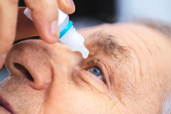
Topical therapy has historically been the first-line treatment for glaucoma management. But with great success come struggles. The most frequent problems with this form of treatment include drop instillation, adherence, cost of medication, and ocular surface disease (OSD). Patients struggling with motor function or coordination can have trouble instilling drops.
In a review by Davis et al, 37% of patients failed to take their first drop.2 Consistently missing this first drop means the medication does not reach the eye and take effect. Further, the medication is wasted.
Medication adherence is an issue, especially in the early stages of glaucoma. Most patients are asymptomatic, so they can often lose faith in the treatment. As more topical therapy is added, the likelihood of adherence decreases. In a study by Friedman et al, 14,000 patients were observed over 1 year, with only 10% found to be fully adherent to medications.3
Cost
The cost of medications can also be a barrier to adequate treatment. Although there are studies that report better efficacy with brand-name medications compared with generics, brand-name medications can cost up to 44% higher than generics.4
OSD is common with glaucoma drops, especially when patients take multiple drops. Preservatives allow the medication to maintain durability, but patients can develop sensitivities. Benzalkonium chloride (BAK) is regarded as one of the most common and most toxic preservatives for eye drops.
With around 70% of eye drops containing BAK, preservatives alter integrity of the cornea.5 In a study by Eamon et al, an estimated 60% of patients taking glaucoma drops exhibited at least 1 symptom of OSD.6 An unhealthy ocular surface can lead to blurry vision, irritation, discomfort, and adherence issues.
Resolving challenges
As clinicians, we must know how to get around complications with drops and find solutions for our patients. Instillation problems can be reduced by demonstrating and guiding patients on proper technique, leading to greater success.
Innovations like the Nanodropper (Nanodropper, Inc) can help keep medication waste to a minimum. The Nanodropper is a bottle attachment that releases a precise amount of medication, reducing overflow and preventing waste.7
Assistance at home can also be beneficial, with a loved one helping not only with instillation but maintaining adherence. Setting alarms on a phone can remind forgetful patients of when to take their drops and improve adherence.
Additionally, there are advancements in dissolvable ocular implants that allow sustained medication release. There is only 1 FDA-approved medication: a bimatoprost intracameral implant (Durysta; Allergan).8
Patients with little or no coverage can take advantage of resources like GoodRx to help with medication costs. Most major classes of drugs have preservative-free formulations, which can be ordered through certain online pharmacies or your local pharmacy. Along with preservative-free options, consider dry eye therapy such as immunomodulators, preservative-free artificial tears, in-office meibomian gland dysfunction treatment options, or punctal plugs.
Glaucoma treatment has evolved over the years to include treatments outside of topical therapy, such as selective laser trabeculoplasty (SLT), angle-based procedures, and filtering devices. These treatment methods have been effective solutions for patients who have failed topical therapy. However, they come with their own difficulties.
SLT
Growing in popularity as a first-line therapy, SLT utilizes photothermolysis to target pigmented cells of the TM, leading to structural changes within the TM that allow for increased aqueous outflow. SLT has few postoperative complications, with the most common being ocular inflammation followed by pain, discomfort, redness, and an increase in IOP.9 These complications are rare and typically only require short-term treatment.
FDA-approved topical steroids were created specifically for treating postoperative inflammation. However, pain, discomfort, and redness typically subside and do not require aggressive treatment.10
Minimally invasive glaucoma surgeries
Minimally invasive glaucoma surgeries (MIGS) have been increasing in use due to their efficacy, safety, and quick recovery time. MIGS include trabecular microbypass stents, Schlemm canal stents, canaloplasty, viscodilation, goniotomy, and subconjunctival stents.
Although there are no major complications associated with stents, occlusion or malposition have been reported in 2.6% to 18% of cases.11 Canaloplasty—which involves insertion of a microcatheter into the TM to facilitate better outflow of aqueous—also has few serious complications. The most common complication after canaloplasty is microhyphema,11 although the presence of a hyphema after canaloplasty can indicate the procedure was successful because access to the drainage system has occurred.
Increase in IOP also occurs in up to 30% of cases but stabilizes 1 to 2 days after the procedure.12 Goniotomies, where a portion of the TM is removed to allow better aqueous outflow, can result in hyphema as well. One study found the rate of hyphema to be 35%; it typically resolved without treatment.13
Lastly, the subconjunctival stent is a newer method of MIGS, in which a gel stent is placed using either an ab interno or ab externo surgical approach. The stent size and shape are designed to reduce hypotony, using principles of laminar fluid dynamics.14
The most common complication of subconjunctival stent placement is occlusion by scar tissue or conjunctiva (32.3%), where reneedling is necessary.15 Although the stent is designed to reduce hypotony, there can be a 24.6% rate of occurrence, according to a study by Grover et al.15
In the same study, 21.5% of patients experienced an increase in IOP greater than 10 mm Hg from baseline.15 Fortunately, postoperative complications with MIGS are mild and require minimal intervention.
Tube shunts
Trabeculectomies and tube shunts are often reserved for severe cases of glaucoma and after angle-based procedures have been attempted. Although highly effective, they are the most invasive of the glaucoma procedures and can be uncomfortable for patients.
According to the Tube Versus Trabeculectomy (TVT) study (NCT00306852), the most common reason for trabeculectomy failure was inadequate IOP lowering (40%), followed by persistent hypotony (31%).16 For tubes, inadequate IOP lowering was also the most common reason for failure in this study, about 54%.16
In the tube group, 29% of patients required additional surgery to lower IOP (26% for trabeculectomies).Furthermore, the rate of hypotony in the tube group was lower than the trabeculectomy group, at 13%.16
Other postoperative complications for tubes include diplopia/strabismus (1.4%-77%), corneal endothelial disease (2%-12%), and a need for bleb or tube revision (10% in the first 6 months).17,18 Postoperative complications from a tube or trabeculectomy should be comanaged with the surgeon who performed the surgery.
Conclusion
Glaucoma is difficult to manage, because every patient has different needs for their type and level of glaucoma. The ultimate goal in glaucoma management is preserving vision while maintaining safety, efficacy, and quality of life. However, the latter tends to be lower on the list of priorities when clinicians get caught up in monitoring IOP and visual fields.
Research has shown that patients with glaucoma tend to have higher incidences of anxiety and depression.19 As health care providers, optometrists should not shy away from the subject of mental health and making sure patients are seeking appropriate care when necessary.
We need to make sure our patients and their families understand the disease, its course, and the need for continued management—a better understanding of the disease can help mitigate anxiety. Additionally, make sure you have low-vision resources available to patients, so they are able to maximize their remaining vision.
REFERENCES
1. Glaucoma awareness can help save vision for millions. National Eye Institute. December 22, 2015. Accessed September 1, 2022. https://www.nei.nih.gov/about/news-and-events/news/glaucoma-awareness-can-help-save-vision-millions
2. Davis SA, Sleath B, Carpenter DM, Blalock SJ, Muir KW, Budenz DL. Drop instillation and glaucoma. Curr Opin Ophthalmol. 2018;29(2):171-177. doi:10.1097/ICU.0000000000000451
3. Friedman DS, Quigley HA, Gelb L, et al. Using pharmacy claims data to study adherence to glaucoma medications: methodology and findings of the Glaucoma Adherence and Persistency Study (GAPS). Invest Ophthalmol Vis Sci. 2007;48(11):5052-5057. doi:10.1167/iovs.07-0290
4. Malvankar-Mehta MS, Feng L, Hutnik CM. North American cost analysis of brand name versus generic drugs for the treatment of glaucoma. Clinicoecon Outcomes Res. 2019;11:789-798. doi:10.2147/CEOR.S156558
5. Steven DW, Alaghband P, Lim KS. Preservatives in glaucoma medication. Br J Ophthalmol. 2018;102(11):1497-1503. doi:10.1136/bjophthalmol-2017-311544
6. Leung EW, Medeiros FA, Weinreb RN. Prevalence of ocular surface disease in glaucoma patients. J Glaucoma. 2008;17(5):350-355. doi:10.1097/IJG.0b013e31815c5f4f
7. Nanodropper data. Nanodropper.com. Accessed September 1, 2022. https://nanodropper.com/data/
8. Shirley M. Bimatoprost implant: first approval. Drugs Aging. 2020;37(6):457-462.
doi:10.1007/s40266-020-00769-8
9. Jha B, Bhartiya S, Sharma R, Arora T, Dada T. Selective laser trabeculoplasty: an overview. J Curr Glaucoma Pract. 2012;6(2):79-90. doi:10.5005/jp-journals-10008-1111
10. Song J. Complications of selective laser trabeculoplasty: a review. Clin Ophthalmol. 2016;10:137-143. doi:10.2147/OPTH.S84996
11. Wellik SR, Dale EA. A review of the iStent trabecular micro-bypass stent: safety and efficacy. Clin Ophthalmol. 2015;9:677-684. doi:10.2147/OPTH.S57217
12. Riva I, Brusini P, Oddone F, Michelessi M, Weinreb RN, Quaranta L. Canaloplasty in the treatment of open-angle glaucoma: a review of patient selection and outcomes. Adv Ther. 2019;36(1):31-43. doi:10.1007/s12325-018-0842-6
13. Salinas L, Chaudhary A, Berdahl JP, et al. Goniotomy using the Kahook Dual Blade in severe and refractory glaucoma: 6-month outcomes. J Glaucoma. 2018;27(10):849-855. doi:10.1097/IJG.0000000000001019
14. Do AT, Parikh H, Panarelli JF. Subconjunctival microinvasive glaucoma surgeries: an update on the Xen gel stent and the PreserFlo MicroShunt. Curr Opin Ophthalmol. 2020;31(2):132-138. doi:10.1097/ICU.0000000000000643
15. Grover DS, Flynn WJ, Bashford KP, et al. Performance and safety of a new ab interno gelatin stent in refractory glaucoma at 12 months. Am J Ophthalmol. 2017;183:25-36. doi:10.1016/j.ajo.2017.07.023
16. Gedde SJ, Schiffman JC, Feuer WJ, Herndon LW, Brandt JD, Budenz DL; Tube versus Trabeculectomy Study Group. Treatment outcomes in the Tube Versus Trabeculectomy (TVT) study after five years of follow-up. Am J Ophthalmol. 2012;153(5):789-803.e2. doi:10.1016/j.ajo.2011.10.026
17. Bailey AK, Sarkisian SR Jr. Complications of tube implants and their management. Curr Opin Ophthalmol. 2014;25(2):148-153. doi:10.1097/ICU.0000000000000034
18. Cardakli N, Friedman DS, Boland MV. Unplanned return to the operating room after tube shunt surgery. Am J Ophthalmol. 2021;229:242-252. doi:10.1016/j.ajo.2021.05.010
19. Quaranta L, Riva I, Gerardi C, Oddone F, Floriani I, Konstas AGP. Quality of life in glaucoma: a review of the literature. Adv Ther. 2016;33(6):959-981. doi:10.1007/s12325-016-0333-6
Articles in this issue
about 3 years ago
Making artificial tears less artificialabout 3 years ago
Orthokeratology is key to managing pediatric myopiaabout 3 years ago
Agent improves near vision for irregular corneas with pinhole effectabout 3 years ago
Refractive technologies encompass a rapidly changing landscapeabout 3 years ago
Dacryostenosis illustrates the complexity of treating teary eyesabout 3 years ago
Q&A: Exploring telehealth through DigitalOptometricsabout 3 years ago
Case study: Pigment epithelial detachment is observed, managedabout 3 years ago
Debunking common ortho-k mythsNewsletter
Want more insights like this? Subscribe to Optometry Times and get clinical pearls and practice tips delivered straight to your inbox.


