Have a patient with diabetes? Get the whole story
Case history can give clues to a patient’s risk of developing diabetic eye disease.
Case history is paramount for gauging the likelihood of finding diabetic eye disease and reducing risk through patient education. On a percentage basis, type 1 diabetes (T1D) confers higher risk of retinopathy after 5 years than does type 2 diabetes (T2D) of any duration, and this risk increases over time.1 That said, recent analysis shows nearly 2% of patients with T2D develop proliferative diabetic retinopathy after 5 years, with a risk 3.5 times higher in patients with T2D who use insulin.2
Insulin use doesn’t mean a patient with T2D has developed T1D but rather that patients with T2D become increasingly insulin deficient over time. Although increasing disease duration is a risk factor for developing and worsening diabetic retinopathy (DR), roughly half of patients with T1D in the Joslin Medalist study (NCT04409795) who had T1D for 50 years or more had no or minimal DR, suggesting longer disease duration in patients without significant DR implies protective genetics in these patients.3
After duration of diabetes, blood glucose control has been considered the second-most important risk factor for DR. Indeed, results of randomized trials and observational studies consistently show elevated glycated hemoglobin A1C (HbA1c) levels predict incident and worsening DR. However, results from the vaunted Diabetes Control and Complications Trial (NCT00360815) show HbA1c levels accounted for a mere 6% to 11% of the total risk for developing DR during the trial.4
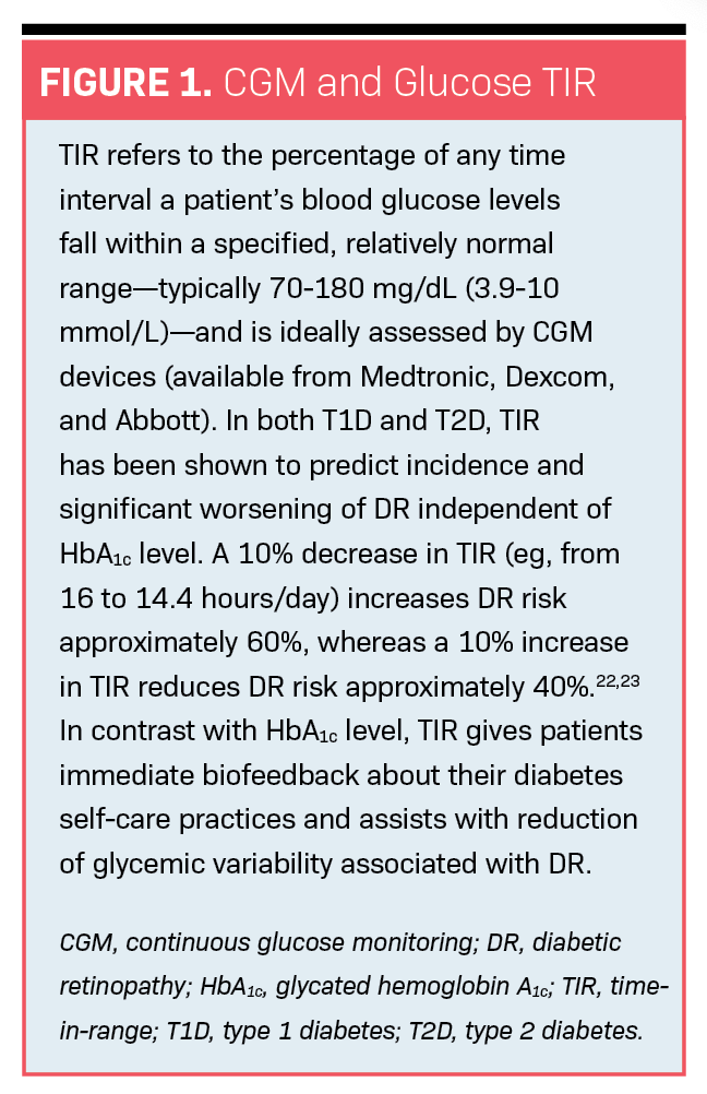
Moreover, identical HbA1c levels from different patients can be associated with different mean blood glucose levels as measured by gold-standard continuous glucose monitoring (CGM) devices. This means a patient with an HbA1c level of 9% may have a better mean glucose level than a patient with an HbA1c level of 7%.5 This discrepancy and other deficiencies of HbA1c level (eg, its value is weighted to the past 2 weeks’ glucose levels before sample collection, and it does not reflect blood glucose variability linked to microvascular complications such as DR) have led to adoption of additional glucose metrics best accessed through CGM, including glucose time-in-range (TIR), which predicts DR independently of HbA1c level (Figure 1). This is not to say HbA1c level is of no value; however, it isn’t the gold-standard metric of diabetes control it has long been considered.
A patient’s current HbA1c level is of little value to clinicians except as it suggests level of control or if it has precipitously decreased within the year (the latter is associated with heightened risk of diabetic reentry retinopathy, ie, DR that develops or worsens with blood glucose levels reentering a normal range—typically a drop of 2 or more points in HbA1c level.)6 It is far more useful to ask patients about their HbA1c level history, as every major prospective diabetes study has shown a metabolic memory phenomenon.7 This refers to the long-term protective/detrimental effects of good/poor glucose control in the first 5 to 10 years after diabetes diagnosis that confer decreased/increased risk of DR over time, despite worsening/improving glycemic control over time.
If a patient has an HbA1c level of 6% now but had poor metabolic control in the 15 years after diagnosis (eg, HbA1c ranging from 8%-12%), we should not be surprised to discover severe DR. In fact, results of recent prospective trials of anti-VEGF therapy in severe nonproliferative diabetic retinopathy found good current glycemic control at study entrance was not at all protective against development of sight-threatening diabetic retinopathy.8 By contrast, severe hypoglycemia itself was linked to higher risk for vision loss in the Fremantle Diabetes Study Phase II,9 possibly a reflection of glycemic variability (ie, patients hospitalized for low blood glucose levels are more likely to have a history of high blood glucose levels) or the fact that hypoglycemia is linked to apoptosis of retinal cells.10
In addition to glucose control, control of blood pressure and lipids, management of obstructive sleep apnea, and absence of nonocular vascular diabetes complications are linked to lower risk of DR, whereas their opposites are associated with higher risk.11,12 Common medications for managing diabetes are also linked to lower risk, including metformin, angiotensin-converting enzyme inhibitors, angiotensin receptor blockers, and statins and fenofibrate.13-16 These medications may be recommended to patients and other health care providers treating them. Patients should be asked whether they consistently take prescribed medications. If they don’t, clinicians should ask why they don’t.
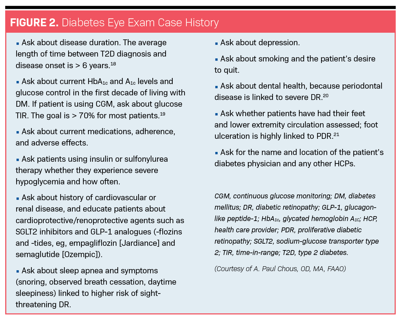
For instance, patients who frequently experience gastrointestinal distress with metformin may find relief with extended-release formulations or by talking to prescribers about alternatives. Taking a patient’s case history is also a great opportunity to ask about prior adherence to annual or biannual dilated eye examinations and puts optometrists in a position to recommend frequent future exams pending diagnostic findings. Patients with a history of depression, renal disease, lower extremity amputation, low education, and low income are at increased risk of being lost to follow-up.17 Optometrists should assiduously counsel, set appointments for, and remind these patients about the importance of ongoing eye care despite the absence of visual symptoms. Figure 2 presents a rational approach to taking a case history in patients with diabetes or prediabetes.
References
1. Yau JWY, Rogers SL, Kawasaki R, et al. Global prevalence and major risk factors of diabetic retinopathy. Diabetes Care. 2012;35(3):556-564. doi:10.2337/dc11-1909
2. Gange WS, Lopez J, Xu BY, Lung K, Seabury SA, Toy BC. Incidence of proliferative diabetic retinopathy and other neovascular sequelae at 5 years following diagnosis of type 2 diabetes. Diabetes Care. 2021;44(11):2518-2526. doi:10.2337/dc21-0228
3. Sun JK, Keenan HA, Cavallerano JD, et al. Protection from retinopathy and other complications in patients with type 1 diabetes of extreme duration: the Joslin 50-year Medalist Study. Diabetes Care. 2011;34(4):968-974. doi:10.2337/dc10-1675
4. Hirsch IB, Brownlee M. Beyond hemoglobin A1c—need for additional markers of risk for diabetic microvascular complications. JAMA. 2010;303(22):2291-2292. doi:10.1001/jama.2010.785
5. Beck RW, Connor CG, Mullen DM, Wesley DM, Bergenstal RM. The fallacy of average: how using HbA1c alone to assess glycemic control can be misleading. Diabetes Care. 2017;40(8):994-999. doi:10.2337/dc17-0636
6. Jingi AM, Tankeu AT, Ateba NA, Noubiap JJ. Mechanism of worsening diabetic retinopathy with rapid lowering of blood glucose: the synergistic hypothesis. BMC Endocr Disord. 2017;17(1):63. doi:10.1186/s12902-017-0213-3
7. Testa R, Bonfigli AR, Prattichizzo F, La Sala L, De Nigris V, Ceriello A. The “metabolic memory” theory and the early treatment of hyperglycemia in prevention of diabetic complications. Nutrients. 2017;9(5):437. doi:10.3390/nu9050437
8. Brown DM, Wykoff CC, Boyer D, et al. Evaluation of intravitreal aflibercept for the treatment of severe nonproliferative diabetic retinopathy: results from the PANORAMA randomized clinical trial. JAMA Ophthalmol. 2021;139(9):946-955. doi:10.1001/jamaophthalmol.2021.2809
9. Drinkwater JJ, Davis TME, Davis WA. Incidence and predictors of vision loss complicating type 2 diabetes: the Fremantle Diabetes Study Phase II. J Diabetes Complications. 2020;34(6):107560. doi:10.1016/j.jdiacomp.2020.107560
10. Emery M, Schorderet DF, Roduit R. Acute hypoglycemia induces retinal cell death in mouse. PLoS One. 2011;6(6):e21586. doi:10.1371/journal.pone.0021586
11. Anwar SB, Asif N, Naqvi SAH, Malik S. Evaluation of multiple risk factors involved in the development of diabetic retinopathy. Pak J Med Sci. 2019;35(1):156-160. doi:10.12669/pjms.35.1.279
12. Smith JP, Cyr LG, Dowd LK, Duchin KS, Lenihan PA, Sprague J. The Veterans Affairs continuous positive airway pressure use and diabetic retinopathy study. Optom Vis Sci. 2019;96(11):874-878. doi:10.1097/OPX.0000000000001446
13. Fan YP, Wu CT, Lin JL, et al. Metformin treatment is associated with a decreased risk of nonproliferative diabetic retinopathy in patients with type 2 diabetes mellitus: a population-based cohort study. J Diabetes Res. 2020;2020:9161039. doi:10.1155/2020/9161039
14. Wang B, Wang F, Zhang Y, et al. Effects of RAS inhibitors on diabetic retinopathy: a systematic review and meta-analysis. Lancet Diabetes Endocrinol. 2015;3(4):263-274. doi:10.1016/S2213-8587(14)70256-6
15. Kang EYC, Chen TH, Garg SJ, et al. Association of statin therapy with prevention of vision-threatening diabetic retinopathy. JAMA Ophthalmol. 2019;137(4):363-371. doi:10.1001/jamaophthalmol.2018.6399
16. Meer E, Bavinger JC, Yu Y, VanderBeek BL. Association of fenofibrate use and the risk of progression to vision-threatening diabetic retinopathy. JAMA Ophthalmol. 2022;140(5):529-532. doi:10.1001/jamaophthalmol.2022.0633
17. Chous AP. How ODs can address patients lost to follow up. Optometry Times®. 2021. Accessed April 24, 2022. https://www.optometrytimes.com/view/how-ods-can-address-patients-lost-to-follow-up
18. Porta M, Curletto G, Cipullo D, et al. Estimating the delay between onset and diagnosis of type 2 diabetes from the time course of retinopathy prevalence. Diabetes Care. 2014;37(6):1668-1674. doi:10.2337/dc13-2101
19. CGM & time in range. American Diabetes Association. Accessed July 3, 2022. https://www.diabetes.org/tools-support/devices-technology/cgm-time-in-range
20. Tandon A, Kamath YS, Gopalkrishna P, et al. The association between diabetic retinopathy and periodontal disease. Saudi J Ophthalmol. 2021;34(3):167-170. doi:10.4103/1319-4534.310412
21. Hwang DJ, Lee KM, Park MS, et al. Association between diabetic foot ulcer and diabetic retinopathy. PLoS One. 2017;12(4):e0175270. doi:10.1371/journal.pone.0175270
22. Beck RW, Bergenstal RM, Riddlesworth TD, et al. Validation of time in range as an outcome measure for diabetes clinical trials. Diabetes Care. 2019;42(3):400-405. doi:10.2337/dc18-1444
23. Lu J, Ma X, Zhou J, et al. Association of time in range, as assessed by continuous glucose monitoring, with diabetic retinopathy in type 2 diabetes. Diabetes Care. 2018;41(11):2370-2376. doi:10.2337/dc18-1131
Newsletter
Want more insights like this? Subscribe to Optometry Times and get clinical pearls and practice tips delivered straight to your inbox.


.png&w=3840&q=75)


