How to address dry eye in the challenging cornea
Understand dry eye’s various factors and causes to get ahead of the treatment curve


My eyes are getting watery after logging off my tenth Zoom meeting this week, which makes me think of the others Zooming away and experiencing dry eye symptoms. As an optometrist who sees corneal calamities on a continual basis, it can become a challenge to treat.
At first, clinicians may diagnose dry eye disease (DED) by observing simple superficial punctate keratitis at the slit lamp. However, complications may ensue, such as recurrent epithelial erosions or persistent epithelial defects, causing more problems. These challenges could lead to neurotrophic ulcers and become even more difficult to handle. Of course, the ideal situation would be to treat only 1 problem at a time—and in a timely manner—so that the cornea improves more quickly and stays healthier longer.
Ocular surface disease can be divided into 2 categories: aqueous deficiency leading to inflammation and lipid deficiency causing exposure keratopathy. These categories can be further split into the component layers of the tear film: The oily coating may be scant due to poorly functioning meibomian glands, or the aqueous layer may be faulty because of an inadequately performing lacrimal gland.
ODs who are able to recognize each element, dissect each part, and understand that the problems can exist alone or together are already ahead of the curve. Knowing this will help them to manage the cornea in a systematic way.
Diagnostic tests
Subjective and objective diagnostic testing can help detect and quantify DED and are easy to do.
Subjectively, surveys can measure patients’ baseline symptoms prior to the visit. One example, the Dry Eye Questionnaire-5, is a quick and effective 5-question test to discover if a patient suffers from aqueous and/or lipid deficiency.
Objective tests that can be executed in clinical practice include matrix metallopeptidases (MMP), tear break-up time, Schirmer’s or phenol red thread test, and slit-lamp evaluation of tear lake meniscus. I prefer the MMP-9 test (InflammaDry; Quidel) for its targeting in determining levels of the inflammatory component in dry eye. Accurately diagnosing and following inflammation allow me to better tailor a management approach.
Levels of severity
For everyone who has been stuck at home binge watching Netflix, you know exactly what it feels like to have dry eyes. For the sake of simplicity, dry eye symptoms can be reduced to the levels of mild, moderate, or severe.
For mild cases, preservative-free artificial tears, such as Refresh Relieva (Allergan) and Retaine MGD (OCuSOFT), are excellent options. Refresh Relieva contains an inactive ingredient, hyaluronic acid, that I have personally and anecdotally found to soothe the corneal surface. For moderate conditions, twice-daily use of Retaine MGD drops combined with a clinically validated nutritional supplement can provide relief to the deficient lipid profile. For severe symptoms, additional therapies should be implemented to maintain the corneal integrity.
Use a supplement
A supplement such as HydroEye (ScienceBased Health) assists with tear film support and contains omega fatty acids that help to prevent the deterioration of the tear film. The combination of γ-linolenic acid (GLA) with eicosapentaenoic acid and docosahexaenoic acid aids in blocking the formation of proinflammatory prostaglandins while simultaneously stimulating production of anti-inflammatory prostaglandins.1
GLA works through its metabolite, dihomogamma linoleic acid to stimulate the tear-specific prostaglandin E1, which reduces inflammation and supports tear production.2 Thus, this complementary therapy can help reduce inflammation and maintain corneal smoothness while improving symptoms of DED.3
Tamp down inflammation
Short-term use of topical steroids can also reduce active surface inflammation. Tapering steroids may be warranted to prevent a possible rebound effect or spike in intraocular pressure.
Kala Pharmaceuticals recently FDA approval for Eysuvis (loteprednol etabonate ophthalmic suspension) for the short-term treatment of the signs and symptoms of DED. Eysuvis incorporates mucus-penetrating particle drug delivery technology to enhnce penetration into target tissues of the eye to suppress inflammation.4
Steroid-sparing anti-inflammatory therapies such as cyclosporine 0.05 (Restasis; Allergan), cyclosporine 0.09% (Cequa; Sun Ophthalmics), and lifitegrast (Xiidra; Novartis) are complementary options that help achieve this goal.
Supplement plus treatment
The use of topical therapies alone may not be sufficient to address corneal deterioration as the severity of ocular surface disease progresses. A cryopreserved amniotic membrane (CAM) such as Prokera (Bio-Tissue) might help. The practice of adding a supplement to work internally while utilizing a CAM to work externally on the corneal surface may enhance recovery time.
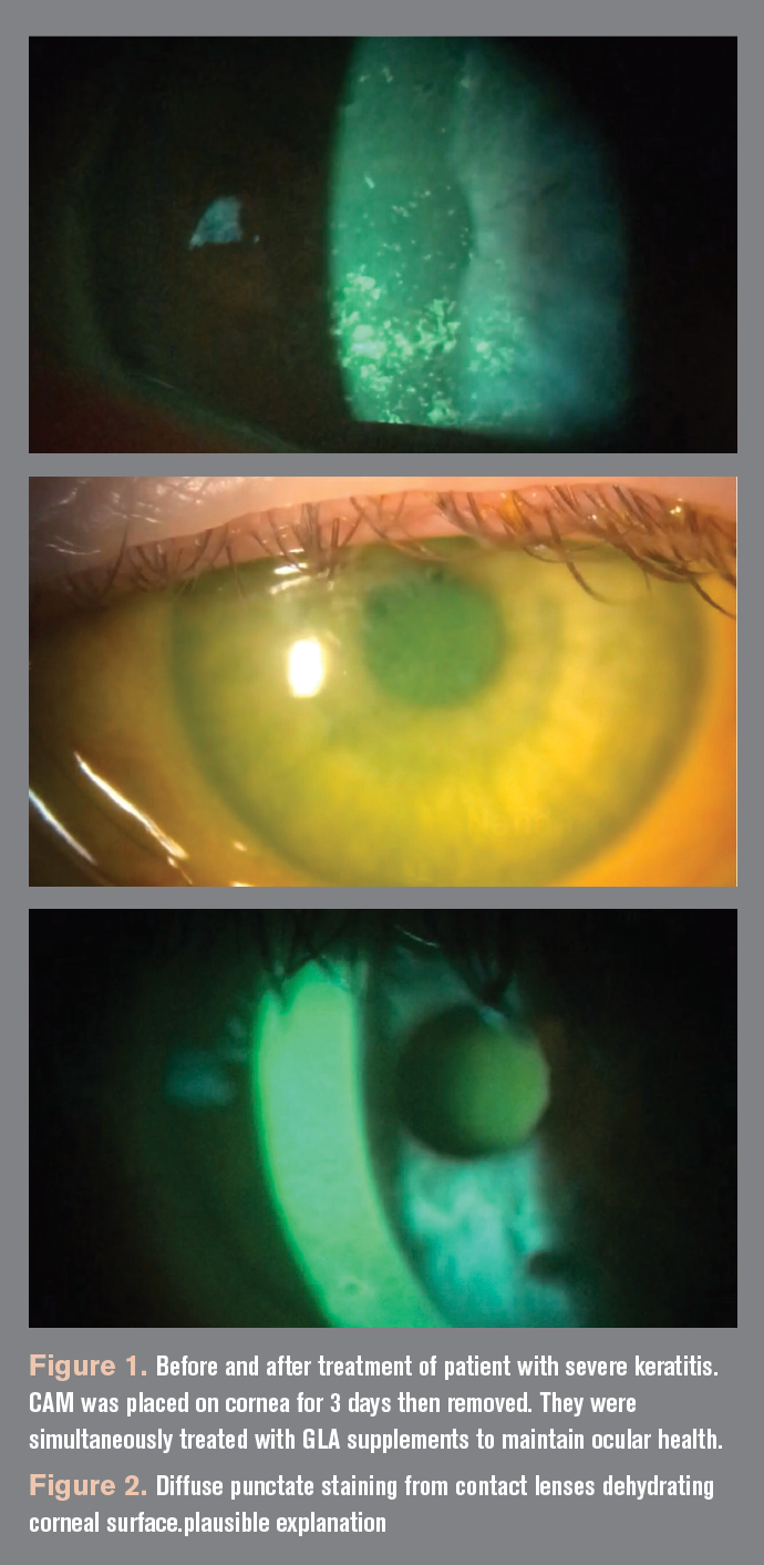
I prefer Prokera as an early treatment option for many corneal disorders due to its healing properties. When used with GLA, a CAM can prevent further deterioration of the ocular surface that may lead to persistent epithelial defects or neurotrophic keratitis. I personally witnessed this renewal in 3 peri-menopausal female patients of mine who all had undergone LASIK surgery several years prior. They all showed moderate diffuse corneal punctate coalescing fluorescein staining which correlated with their symptoms of moderate to severe dry eyes.5 Therefore, in my clinic, I insert the CAM and balance it with the oral consumption of HydroEye twice a day.
All 3 individuals noticed dramatic improvement in their ocular signs with resolution of punctate erosions occurring within 1 week. Typically, healing time for these lesions may take 3 months or longer, depending on severity. Corneal restoration with Prokera is accelerated from a period of several months to a few days, even in recalcitrant cases of keratitis.
Contact lens conundrums
Contact lenses can also be a challenge in persons with corneal concerns. Occasionally, busy practitioners can become too focused on contact lens modalities but forget to truly evaluate the patients’ dry eye complaints. A classic example is the patient who routinely uses multiple digital devices simultaneously, concentrating on a tablet and smart phone while watching a big-screen TV. Subsequently, their vision blurs as their contact lenses fall onto their keyboard. The patient blames the contact lenses instead of device usage.
Because eyecare professionals frequently think of the lens design or material as causing the problem, they may not consider the lacrimal or meibomian glands as possible culprits. One should assess for inflammation and then express the meibum to see the quality of the lipid. Obviously, if it is clear and serous as olive oil there is no problem. However, if the expressed lipid looks thick and gelatinous as lard, then I immediately start HydroEye therapy and may recommend insertion of Prokera. The patient will notice a significant decrease in their dry eye symptoms within a few weeks and their contacts will actually stay in.
Deal with pressure
Treating glaucoma can be unnerving because topical medications to lower the intraocular pressure can contribute to the cornea’s decline. Most of these drugs have preservatives that can be toxic to the corneal epithelium over prolonged use. Therefore, if a patient with dry eye is also on a prostaglandin therapy, one should switch to a preservative-free or benzalkonium chloride-free option such as tafluprost (Zioptan; Akorn) or latanoprost (Xelpros, Sun Ophthalmics). Another cutting-edge possibility is an innovative sustained-release intracameral injection of bimatoprost implant (Durysta; Allergan). With the offending preservative removed, a cryopreserved amniotic membrane with a quality GLA supplement can improve the ocular surface as the patient continues their pressure lowering medications.
Follow-up
I advise my patients to return to the clinic at close intervals until they are stable and then extend visits out as conditions improve. In my experience, patients who do not return for regular maintenance check-ups and follow-up visits may be at greater risk of breaking down. Thankfully, multiple therapies are now available to treat challenging corneal cases. Once these innovative approaches are employed, both the patients and practitioners will be happier with their outcomes and then they can watch as many movies as they like.
References
1. Barham JB, Edens MB, Fonteh AN, Johnson MM, Easter JL, Chilton FH. Addition of eicosapentaenoic acid to gamma-linolenic acid-supplemented diets prevents serum arachidonic acid accumulation in humans. J Nutr. 2000;130(8):1925-1931. doi: 10.1093/jn/130.8.1925
2. Brignole-Baudouin F, Baudouin C, Aragona P, et al. A multi-centre, double-masked, randomized, controlled trial assessing the effect of oral supplementation of omega-3 and omega-6 fatty acids on a conjunctival inflammatory marker in dry eye patients. Acta Ophthalmol. 2011;89(7):e591-e597. doi: 10.1111/j.1755- 3768.2011.02196.x
3. Sheppard JD Jr, Singh R, McClellan AJ, et al. Long-term supplementation with n-6 and n-3 PUFAs improves moderate-to-severe keratoconjunctivitis sicca: a randomized double-blind clinical trial. Cornea. 2013;32(10):1297-304. doi: 10.1097/ ICO.0b013e318299549c
4. Eysuvis. Prescribing information. Kala Pharmaceuticals; 2020. Accessed January 19, 2020. https://www.accessdata.fda.gov/ drugsatfda_docs/label/2020/210933s000lbl.pdf
5. Thomas J, Tighe S, Sheha H, et al. Corneal nerve regeneration after self-retained cryopreserved amniotic membrane in dry eye disease. J Ophthalmol. 2017;2017:6404918. doi: 10.1155/2017/6404918

Newsletter
Want more insights like this? Subscribe to Optometry Times and get clinical pearls and practice tips delivered straight to your inbox.