Technology helps to diagnose corneal ectasia
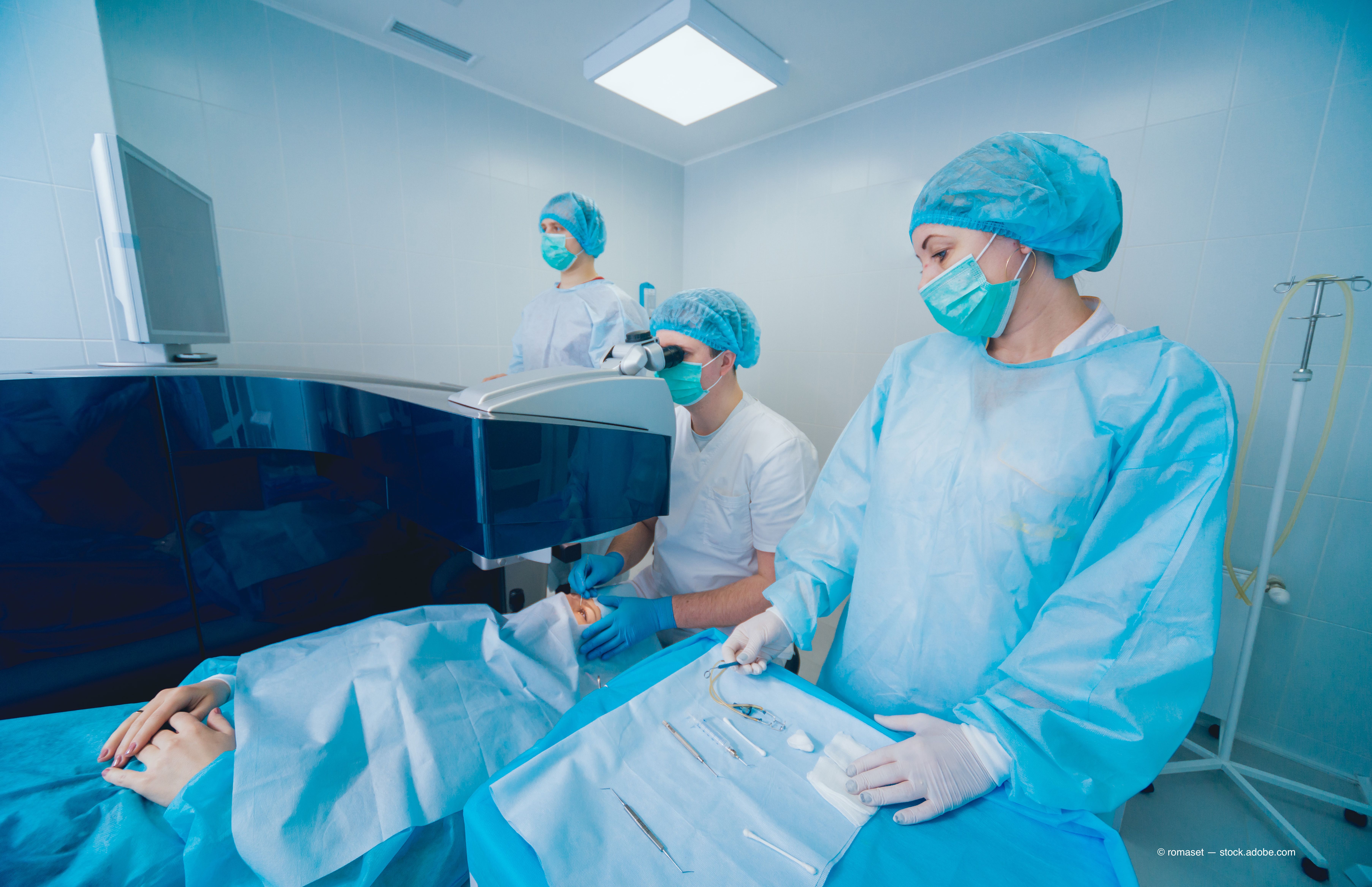
Corneal ectasia is one of the worst outcomes of laser eye surgery (LASIK). One OD takes a look at the importance of operating on good candidates and the newest technologies to do so succesfully.


Almost two decades ago, “The Cornea is not a Piece of Plastic” was published, and it outlined how Munnerlyn’s formula is the appropriate starting point for evaluating how much tissue is removed from the cornea and how it responds. Unfortunately, it does not answer the most important question, what is the biomechanical stability of the cornea.1
The challenge is the cornea is not plastic but visco-elastic, having properties of both a viscous and elastic media.
One of the worst outcomes of laser refractive surgery is corneal ectasia, and the ability to diagnose this condition has changed over time. An ectatic cornea is often said to lack stiffness, but stiffness is not a biomechanical property. An ectatic cornea is a combination of bending, shear, tensile, and compressive properties of a material along with the geometry of that material.
In 2018, our diagnostic abilities have improved from early placido disc topography and manual pachymetry to corneal tomography that allows us to examine the structure of the cornea.
The cornea does not have linear elastic properties and is best described as hyper-elastic. In this bend-but-don’t-break model, the corneal collagen fibers remain loose during the initial stress-like an uncoiling spring-but tighten after significant stress.3,4
Previously from Dr. Owen: Why I changed what I tell patients about refractive surgery
We have learned not all corneal collagen fibers have the same biomechanical properties. The peripheral cornea is stiffer than the central cornea and circumferentially running fibers because of a circular ligament of fibers.5
Using tomography
Tomography provides data about the relationship of the curvature along with the thickness throughout the cornea. From this, we have learned the importance of the cornea’s back curvature, thickness progression, and thinnest and steepest areas.5
Software such as the Belin/Ambrosio Enhanced Ectasia Display (BAD) on the Oculus Pentacam has been developed to evaluate data from tomography and to provide a more analytical approach.
This software analyzes anterior and posterior elevations, pachymetric progression, and a cornea-thickness special profile to come up with a big “D.” The big “D” is a compilation of nine separate parameters and their standard deviation from a normal population. These parameters include keratometic values, corneal thickness, and progression index. In a study comparing 46 keratoconic eyes to 214 normal eyes, researchers found a 99 percent sensitivity for keratoconus when the big “D” was greater than 2.0 standard deviations from normal.5
ODs believe the measurements we currently evaluate-corneal thickness, corneal elevation, back surface elevation-are effects of a change or altered biomechanical structure. In an effort to identify subclinical keratoconus, it is important to measure the biomechanical properties of the cornea.
Related: Femtosecond advances help ODs, patients
Corneal crosslinking is a procedure that improves the strength of the cornea, but the only outcome measure we currently use is the lack of progression of corneal steepening-the proxy for biomechanical strength.
Corneal hysteresis digs deeper
Corneal hysteresis is a measurement of the visco-elastic properties of the cornea. It can be described as the energy loss of a visco-elastic material between the loading and unloading in a stress-stain analysis.
The theory behind hysteresis makes it seem useful for laser vision correction. Rather than measure the shape and thickness of the cornea, hysteresis is the measurement of the actual ability to respond to stress in a valuable data point.
Ocular Response Analyzer (Reichert, Inc.) uses bidirectional noncontact applanation pneumotonometry to measure hysteresis. This device has shown to be valuable in the diagnosis and monitoring of glaucoma patients. Hysteresis of the cornea can provide a corneal compensated intraocular pressure (IOP), which has been proven to be less influence by corneal properties and a better predictor of visual field loss.6,7
There is a statistically significant positive correlation among each of corneal hysteresis (CH) and corneal resistance factor (CRF) and the central corneal thickness, but the correlation is less for CH (CH, r=0.4655; CRF, r=0.5760). Hysteresis is statistically significantly lower in keratoconic eyes as compared to normal eyes.8,9
Related: How to manage vision changes over time post-LASIK
Corvis ST (Oculus, Inc.) is a device used to measure the biomechanical properties of the cornea, but it is not yet approved for use in the U.S. by the Food and Drug Administration (FDA). Corvis ST uses an air puff to deform the cornea, and a high-speed camera (a Scheimpflug camera) captures the deformation and resulting deflecting.
IOP is a confounding factor in determining corneal biomechanics because as IOP increases, it creates wall of tension, causing the cornea to appear to be stiffer. It is important to create normative data for all of the parameters as a function of age and IOP.
The Vincinguerra Screening Report is the result of a multicenter study of 705 healthy patients to establish normative values for Corvis ST.10 This study evaluated many biomechanical properties including deformation amplitude, maximum corneal inward velocity, maximum corneal outward velocity, and maximum deformation area.
From the Vincinguerra Screening Report, the Corvis Biomechanical Index (CBI) was developed. The CBI is based on a linear regression analysis that has been shown to be both sensitive (94.3 percent) and specific (97.5 percent) in detecting clinical keratoconus.11
Tomographic/Biomechanical Index assists
Having biomechanical normative data and thresholds to identify keratoconus is valuable when screening patients for laser vision correction, but how do these parameters interact with our existing knowledge of corneal curvature and thickness, and are we increasing the sensitivity and, we hope, making laser vision correction a safer procedure?
A software program, Tomographic/Biomechanical Index (TBI) that uses Scheimpflug-based corneal tomographic from Pentacam HR and biomechanical analysis from Corvis ST exams, was developed to address those questions. The TBI cut-off value of 0.79 provided 100 percent sensitivity for detecting clinical ectasia (keratoconus and VAE-E groups) with 100 percent specificity.12
Related: Preparing your patient for PRK
The TBI was sensitive for detecting subclinical (fruste) ectasia among eyes with normal topography in very asymmetric patients. This information becomes valuable in identifying patients at risk and properly communicating that risk to patients considering laser vision correction.
In an unpublished retrospective analysis conducted at TLC Toronto, researchers evaluated 1056 patient records. The BAD was used to evaluate if a patient would be a candidate for Laser eye surgery (LASIK), photorefractive keratectomy (PRK), or no surgery by a trained experience clinician. That decision was compared to a decision that included TBI data.
In the vast majority of patients, the addition of the TBI data did not change the decision on what (Lasik or PRK) or if any procedure should be performed. In 13.2 percent of the cases, patients who had abnormal BAD but normal TBI were asked to change their current procedure. Finally, 3.4 percent of cases with normal BAD but abnormal TBI were converted from LASIK to PRK, or from PRK to no surgery.
Future bright
In 2018, the outcomes from the excimer laser are more precise than ever.13 The ability to make a flap with a femtosecond laser has reduced many complications that existed with the microkeratome.14
Nevertheless, operating on a poor candidate can result in a poor outcome despite technology. As the ability to identify biomechanically stable corneas continues to improve, it should result in a safer, more efficacious procedure.
References
1. Roberts, C. The cornea is not a piece of plastic. J Refract Surg. 2000 Jul-Aug;16(4):407-13.
2. Fratzl P1, Misof K, Zizak I, Rapp G, Amenitsch H, Bernstorff S. Fibrillar structure and mechanical properties of collagen. J Struct Biol. 1998;122(1-2):119-22.
3. Liu X, Wang L, Ji J, Yao W, Wei W, Fan J, Joshi S, Li D, Fan Y. A mechanical model of the cornea considering the crimping morphology of collagen fibrils. Invest Ophthalmol Vis Sci. 2014 Apr 28;55(4):2739-46.
4. Meek KM, Newton RH. Organization of collagen fibrils in the corneal stroma in relation to mechanical properties and surgical practice. J Refract Surg. 1999 Nov-Dec;15(6):695-9.
5. Ambrósio R Jr, Caiado AL, Guerra FP, Louzada R, Sinha RA, Luz A, Dupps WJ, Belin MW.. Novel pachymetric parameters based on corneal tomography for diagnosing keratoconus. J Refract Surg. 2011 Oct;27(10):753-8
6. Medeiros FA, Weinreb RN. Evaluation of the influence of corneal biomechanical properties on intraocular pressure measurements using the ocular response analyzer. J Glaucoma. 2006 Oct;15(5):364-70.
7. Medeiros FA, Meira-Freitas D, Lisboa R, Kuang TM, Zangwill LM, Weinreb RN. Corneal hysteresis as a risk factor for glaucoma progression: a prospective longitudinal study. Ophthalmology. 2013 Aug;120(8):1533-40.
8. Fontes BM, Ambrósio R Jr, Alonso RS, Jardim D, Velarde GC, Nosé W. Corneal biomechanical metrics in eyes with refraction of -19.00 to +9.00 D in healthy Brazilian patients. J Refract Surg. 2008 Nov;24(9):941-5.
9. Shah S, Laiquzzaman M, Bhojwani R, Mantry S, Cunliffe I. Assessment of the biomechanical properties of the cornea with the ocular response analyzer in normal and keratoconic eyes. Invest Ophthalmol Vis Sci. 2007 Jul;48(7):3026-31.
10. Joda AA, Shervin MM, Kook D, Elsheikh A. Development and validation of a correction equation for Corvis tonometry. Comput Methods Biomech Biomed Engin. 2016;19(9):943-53.
11. Vinciguerra R, Ambrósio R Jr, Elsheikh A, Roberts CJ, Lopes B, Morenghi E, Azzolini C, Vinciguerra P. Detection of Keratoconus With a New Biomechanical Index. J Refract Surg. 2016 Dec 1;32(12):803-810.
12. Ambrósio R Jr, Lopes BT, Faria-Correia F, Salomão MQ, Bühren J, Roberts CJ, Elsheikh A, Vinciguerra R, Vinciguerra P. Integration of Scheimpflug-Based Corneal Tomography and Biomechanical Assessments for Enhancing Ectasia Detection. J Refract Surg. 2017 Jul 1;33(7):434-443.
13. ClinicalTrials.gov. Wavefront-guided PRK vs wavefront-optimized PRK. Available at: https://clinicaltrials.gov/ct2/show/NCT02091934. Accessed 4/18/18.
14. Callou TP, Garcia R, Mukai A, Giacomin NT, de Souza RG, Bechara SJ. Advances in femtosecond laser technology. Clin Ophthalmol. 2016 Apr 19;10:697-703.

Newsletter
Want more insights like this? Subscribe to Optometry Times and get clinical pearls and practice tips delivered straight to your inbox.