Welcome to the EVO-lution
Evolving phakic IOL offers high-definition vision.
In January 2004 I published an article marveling at the almost approved, newest refractive surgical procedure. As a subinvestigator in the clinical trials for the only phakic intraocular lens that sat in the posterior pole, I had a front row seat to one of the greatest refractive opportunities of our time.
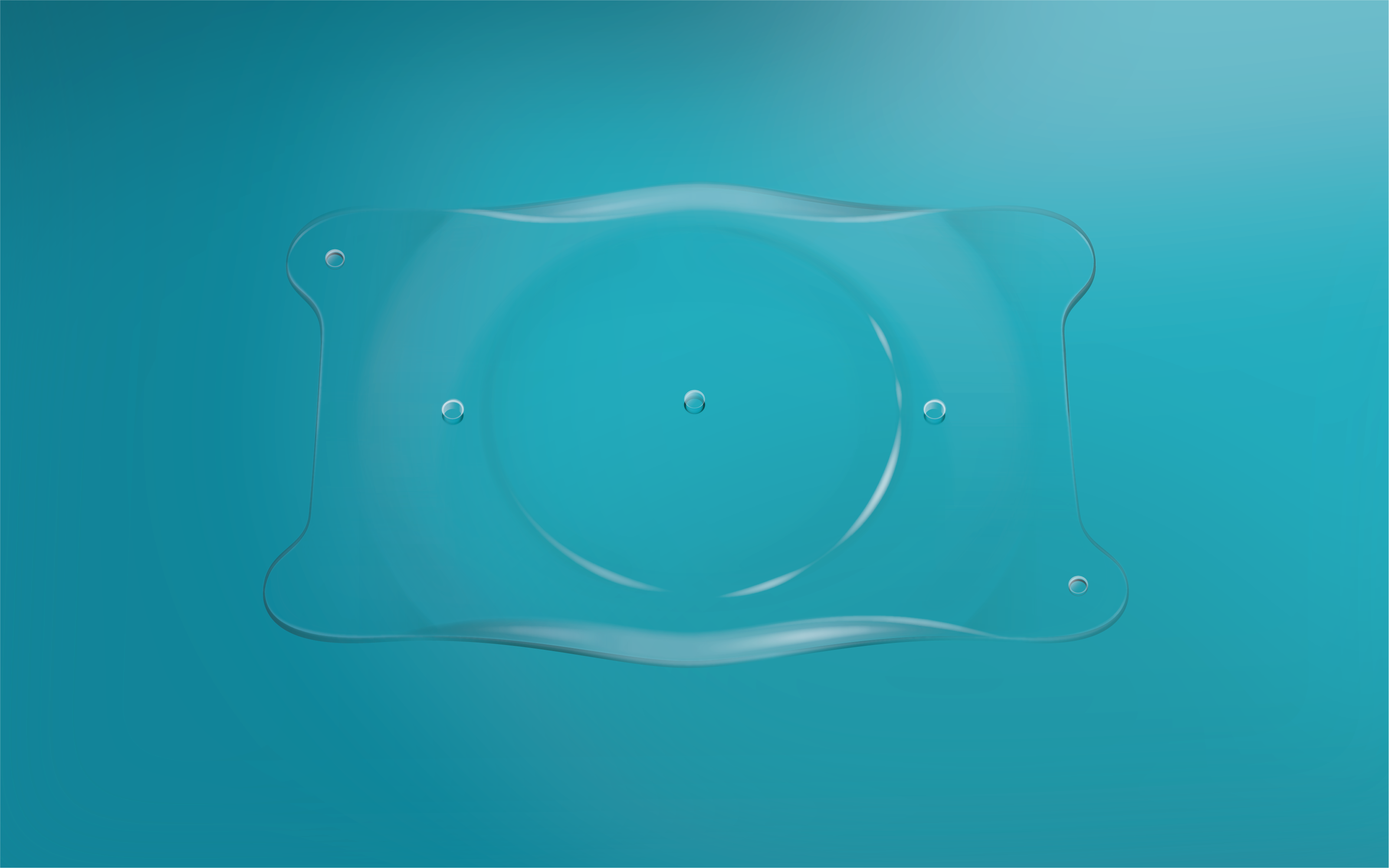
Image courtesy of STAAR Surgical.

STAAR Surgical is the purveyor of this refractive gem, the Visian Intraocular Collamer lens (ICL).
However, unlike the makers of Coca-Cola—who changed the original formula and failed (that new Coke was trash!)—STAAR made 1 small tweak to the ICL and ushered in an evo-lution with this phakic IOL.
Arguably, the ICL should be considered the gold standard for refractive surgery. LASIK often is pseudonymous with all refractive procedures in the eyes of our patients.
Although LASIK has made great strides with the addition of wavefront technology, the use of a femtosecond laser to create a predictable and uniform flap, and a greater understanding of the preoperative parameters for success, it is still limited.
Remember, patients contemplate pursuing refractive surgery for 3 to 7 years before they make their pilgrimage into a surgery center for an evaluation. Notwithstanding the bias that each clinician may harbor regarding their own experiences, and thus place unwanted limitations on their patients.
For someone who has been an active participant, advocate, and cheerleader, I harbor no such bias. There is—with very few exceptions—a refractive option for most of our patients. It just requires matching them with the best option.

When I see a patient with moderate myopia, or a patient with thinner-than-comfortable corneas, or tomographies and topographies that are dubious, I get excited to let them know that we have a much better option than excimer laser.
The patient’s deer-in the-headlight effect is broken by my very straightforward and exuberant discussion about the enhanced visual opportunity we can expect from this remarkable proprietary copolymer material, derived from porcine collagen and a polymer base—aptly named Collamer.
If you are unaware of the phakic IOL, you are missing out on a refractive wunderkind. With correction of myopia in ranges of —3.00D to —20.00D and astigmatism in the range of 1.00D to 4.00D, it ensures that very few patients are denied their desire to be free of glasses or contacts.

With cornea-based procedures, we work at the fringes of the eye’s refractive borders— unlike this phakic lens, which sits as close to the nodal point as possible.
My discussion for almost 20 years has revolved around the ease and success of this lens that behaves like a soft contact lens in the eye, implanted through an incision equivalent to those made millions of times a year by cataract refractive surgeons. The beauty of this procedure is the simplicity and elegance of the placement in the ciliary sulcus.
You may not know that the original phakic IOLs sat in the anterior chamber. This gave surgeons a great view of the placement, which for some of the worst iterations literally clipped onto the iris. I think you can see how an acrylic lens that sits on the iris in the anterior chamber may not have been the best option. Thus, the ICL posterior positioning should be considered ideal. Especially since, without any removal of tissue, worry of pupil size, or possibility of any flap-related issues, and coupled with the fact you could remove the lens, the ICL procedure is still a modern improvement in refractive surgery.
The ICL is to refractive surgery as the iPhone was to cellular technology 15 years ago. Both brought better options and ease of use to those who wanted the technology. Much like Apple, STAAR didn’t just rest on their technology without researching how to create a more enhanced procedure.
When this lens was under FDA investigation in the early 1990s, a drawback was the extra step of creating a peripheral iridectomy, preferably preoperatively assisted with a YAG laser or the more preferred European method of doing it surgically during the ICL implantation process.
Communication is always a key part of the preoperative and postoperative journey. Take, for example, how we discuss dysphotopsia following multifocal IOL surgery. I still tell these patients that they need to look at a light source after surgery with the hope that they will see concentric rings emanating from the source. The proper placement of the lens ensures the patient will see these rings around the light.
I always tell my patients what adverse effects may happen, rather than not telling them and having them be under the false impression these events are unwanted complications. Thus, I have heard from enough patients that the YAG laser peripheral iridotomy (PI) was equivalent to being snapped by a rubber band.
Another unfortunate side effect of the iridotomy is that they were not always large enough or did not have full thickness so that the aqueous transferred easily from the posterior to the anterior chamber; therefore, a rise in IOP was an unfortunate consequence. Treating these patients often meant being diligent around the implantation and the IOP.
Moreover, some surgeons attempted to thwart this complication by enlarging the PIs and creating glare or, in the very rare case, diplopia. This latter option may be the reason why some ICL patients have unwanted dysphotopsia. It has been my experience that patients neuroadapt or that the inevitable chalasis of the lids may obscure the PI, therefore decreasing the unwanted glare. Yet, these patients are younger and that may take more time than your patients are willing to invest. This means more hand holding and chair time, which no one wants.
Which brings us to the EVO!
In 2005, when the ICL V4 was rolling out in the US, investigators questioned how they could avoid the PI. The addition of a 0.36-mm hole in the center of the ICL V4 optic was evaluated for both the transfer of aqueous and continuation of vision.
The EVO ICL has been in use outside the US for more than a decade, with more than 1 million implants worldwide. The EVO is now FDA approved and the standard of care for ICL patients. The availability of the EVO ICL ensures that the preoperative treatment of these patients is as seamless as the postoperative treatment.
As with any refractive surgery, a thorough ocular surface evaluation is a necessity, as well as a discussion about expectations. The postoperative evaluation of the lens should include visualization of the vault, the space from the posterior EVO ICL to the anterior crystalline lens, some mild anterior chamber inflammation, and IOP measurements.
I usually see these patients at 1 day and
1 week postoperatively. A 1-month follow-up is often not necessary because the quality of the patient’s vision and the quiet anterior chamber make this visit superfluous.
The main emphasis during follow-ups is ensuring the toric ICL is in the correct alignment and that the lens has a sufficient vault from the crystalline lens. I still use the thickness of the cornea, via Purkinje images, as my measurement for the vault. Although it is unlikely to see the vault change, you want to know where it is as time moves forward. Without a PI, there is little else to evaluate.
STAAR has big plans for their EVO platform with presbyopic ICLs, giving the hyperopic community another refractive option.
For now, I am just glad that we can avoid the rubber band snap and instead usher our patients straight toward amazing high-definition vision.
Welcome to the EVO-lution.

Newsletter
Want more insights like this? Subscribe to Optometry Times and get clinical pearls and practice tips delivered straight to your inbox.