Coats’ disease: A case study of a 4-year old boy
Early and correct diagnosis of this congenital retinal vasculopathy vital to improve visual outcomes.
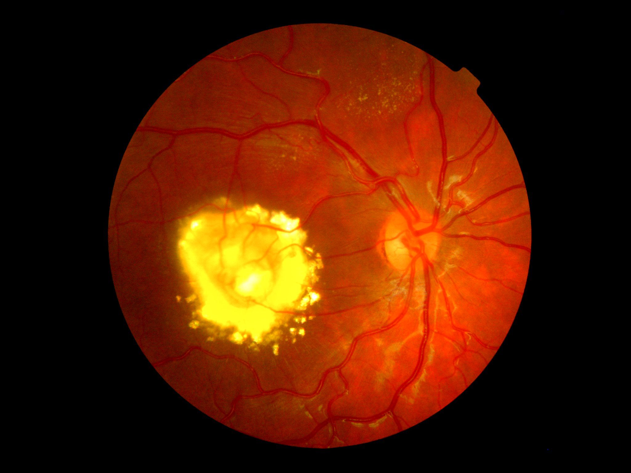
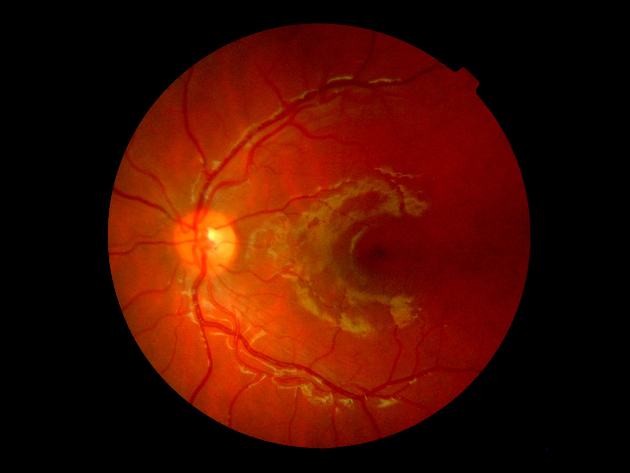
Figure 1. Right and left posterior pole of a 4-year-old patient with suspected Coats disease. Note the subfoveal nodule with associated exudative retinopathy.


This case study highlights the importance of a comprehensive eye examination with pupil dilation early in life, regardless of the presence or absence of signs and/or symptoms of pathology.
Recently, a 4-year-old African American male presented for his first eye examination. As per the child’s mother and father, the chief complaint was that he had failed a vision screening at his pediatrician’s office for the reasons of hyperopia and astigmatism in the right eye.
Observation the child walking back to the examination room showed a normal gait and overtly good attention to his surroundings. Medical history was unremarkable, and he was taking no medications. He also had no known medication or environmental allergies. Family history was remarkable for cataracts, high blood pressure, and glaucoma.
Related: A parent's perspective on genetic testing
Exam details
Entering uncorrected distance visual acuities by means of Allen pictures were 20/400 and 20/25 for the right eye and left eye, respectively. Near visual acuity was 20/20 with both eyes open. Color vision was normal for the left eye and was unable to be tested for the right eye. There was no frank stereopsis present.
The patient was not fully cooperative for cover test assessment, but Hirschberg testing showed no overt strabismus. Pupil testing, extraocular muscle function, and visual field assessment by means of quadrant screening were all unremarkable as well.
Anterior segment examination showed clear corneas, intact irides, open angles, and quiet lids, lashes, and conjunctivae. Anterior chambers were deep and quiet. Intraocular pressure measurements by means of rebound tonometry were 9 mm Hg in the right eye and 10 mm Hg in the left eye.
Related: Offer support to patients with inherited eye diseases
Consent was obtained from the parents for dilation, and wet retinoscopy revealed +0.75 DS in the left eye and a highly variable reflex reading in the right eye ranging from +3.75 DS to +1.00 DS with a small but variable degree of with-the-rule astigmatism.
Posterior pole examination of both eyes is as shown in Figure 1. Note the raised nodular lesion underneath the fovea and the surrounding coalesced exudative retinopathy.
More by Dr. Casella: SD-OCT shows schisis advancements due to sickle cell
Diagnosis
I informed the child’s parents that my initial diagnosis was Coats’ disease and arranged for an urgent evaluation with a retina specialist.
I also explained the guarded visual prognosis for the right eye given foveal involvement and prescribed spectacles with impact-resistant lenses to be worn full-time to protect the left eye. At the time this piece was written, I had not heard the results of the child’s consultation with the retina specialist.
More by Dr. Casella: Be leery of large optic cups when screening for pediatric glaucoma
Discussion
Coats’ disease is a typically congenital retinal vasculopathy characterized by retinal telangiectasia and subsequent exudative retinopathy.1 This condition is non-hereditary and is predominantly unilateral with a predilection for males.2
As Coats’ disease progresses, the potential for poor visual prognosis increases.1 The most severe forms of this condition result in total retinal detachment, neovascular glaucoma, and phthisis bulbi, which are visually devastating and may necessitate enucleation.3
Coats’ disease was first described in 1908 by George Coats as a unilateral vascular disease occurring in the retinas of young males.4 It can have a highly variable clinical appearance based on the severity of the disease process at presentation.
Coats’ disease (along with, among other conditions, cataract, retinoblastoma, and persistent hyperplastic primary vitreous) is one of the causes of leukocoria and/or strabismus in a child.4 This is one reason why patients with this condition may be referred for suspicion of retinoblastoma.
More by Dr. Casella: Why taking patient history never ends
As far as visual function is concerned, one finding which leads to a more negative prognosis is the presence of a subfoveal nodule.1 Unfortunately, this patient has one of these nodules (see Figure 1). The nodules seen in some presentations of Coats’ disease contain fibrotic tissue and unfortunately have a predilection for the macula.5 These subfoveal nodules can be associated with retinal–retinal anastomosis as evidenced by fluorescein angiography;1 telangiectasias and associated areas of nonperfusion can also be evidenced in this way.1
Related: Consider IOP fluctuations when diagnosing glaucoma
Treatment of Coats’ disease is difficult and centers mainly around an attempt to lessen the retina’s demand for vascular growth. As such, laser photocoagulation and anti-vascular endotheliam growth factor (VEGF) therapy have shown efficacy in the earlier disease process, and anti-VEGF therapy with vitrectomy have shown efficacy in advanced disease.6
As with any ocular disease, early and correct diagnosis coupled with timely treatment is key in the hopes of achieving a more favorable ocular and visual outcome. Therefore, Coats’ disease is one of the reasons ODs advocate for comprehensive eye examinations with pupil dilation early in life, regardless of the presence or absence of signs and/or symptoms of pathology.
More by Dr. Casella: Look at more than the optic nerve head in glaucoma patients
References:
1. Daruich AL, Moulin AP, Tran HV, Matet A, Munier FL. SUBFOVEAL NODULE IN COATS' DISEASE: Toward an Updated Classification Predicting Visual Prognosis. Retina. 2017 Aug;37(8):1591-1598.
2. Zhao Q, Peng XY, Yang WL, Li DJ, You QS, Jonas JB. Coats' disease and retrobulbar haemodynamics. Acta Ophthalmol. 2016 Jun;94(4):397-400.
3. Shields JA, Shields CL, Honavar SG, Demirci H, Cater J. Classification and management of Coats disease: the 2000 Proctor Lecture. Am J Ophthalmol. 2001 May;131(5):572-83.
4. Balmer A, Munier F. Differential diagnosis of leukocoria and strabismus, first presenting signs of retinoblastoma. Clin Ophthalmol. 2007 Dec;1(4):431-9.
5. Chang MM, McLean IW, Merritt JC. Coats' disease: a study of 62 histologically confirmed cases. J Pediatr Ophthalmol Strabismus. 1984 Sep-Oct;21(5):163-168.
6. Yang X, Wang C, Su G. Recent advances in the diagnosis and treatment of Coats’ disease. Int Ophthalmol. 2019 Apr;39(4):957-970.
