
Publication|Articles|September 16, 2024
- September digital edition 2024
- Volume 16
- Issue 09
Efficiency of physiotherapy in treating and preventing uncomplicated acquired myopia
An inside look into a retrospective study conducted from 2019 to 2023 that aimed to assess the effectiveness of treatment and prevention of the progression of uncomplicated acquired myopia through physiotherapeutic methods.
Advertisement
Myopia is one of the most common ocular pathologies. The cause of the onset and progression of myopia is not fully understood, but its prevalence is increasing globally. If the number of cases of myopia continues to rise at the current rate, it is estimated that by 2050 approximately 5 billion people will be myopic.1 As a result, myopia warrants national and international collaborative efforts as the cost and public health consequence are massive and frequently underrated in trends.2
A retrospective study
A retrospective study was conducted from 2019 to 2023 in accordance with the principles of the World Medical Association’s Declaration of Helsinki: Ethical Principles for Medical Research Involving Human Subjects. The aim of the study was to assess the effectiveness of treatment and prevention of the progression of uncomplicated acquired myopia through physiotherapeutic methods. It was initiated only after obtaining informed consent from all participants, following an explanation of the applied methods and the consequences of the research.
The study included 80 participants, all of whom were aged 6 to 11 years. These participants were divided into two groups of 40 eyes each: the base group, which underwent physiotherapeutic treatment, and the control group, whose patients used single vision spectacle lenses. Furthermore, the eyes in each group were randomized into 2 subgroups based on the degree of myopia (low or moderate), and these subgroups were further divided into 2 groups, depending on the presence of a familial factor. For the comparative assessment of the dynamics of the studied indices throughout the study, the clinical cases were evaluated before treatment, and after 1 and 2 years of treatment to align with the annual gradient of myopia progression.
Applied treatment methods
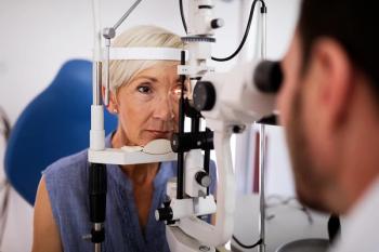
a) Electropuncture: This method aims to stimulate the somato-vegetative functions of the body reflexively by applying transcutaneous electrical current to biologically active points (peripheral reflex zones) (Image 1).
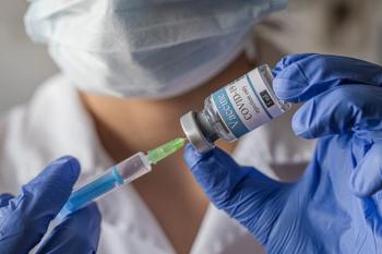
b) Low-intensity laser stimulation of the ciliary muscle: This method involves stimulating the ciliary muscle using low-intensity helium-neon laser radiation, employing infrared laser light. Low-intensity helium-neon laser radiation enhances biosynthetic and proliferative processes in the conjunctival tissue of the ciliary body, thereby improving the vascularization of the ciliary muscle, which positively influences the accommodation reflex (Image 2).
c) Combined treatment: Electropuncture plus low-intensity laser stimulation of the ciliary muscle is a patented method.3
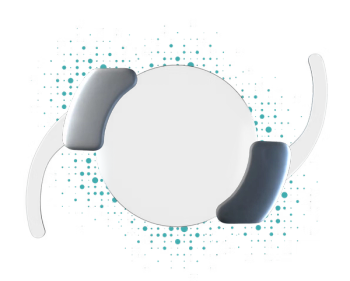
d) Optical-reflex treatment of accommodation: Spherical positive lenses relax the ciliary muscle (Image 3).
Results and discussions
In the base group with low myopia and a family factor, after the first year, the annual myopia progression rate based on the spherical equivalent (AMPRS) value decreased from –0.68 plus or minus 0.05 diopter (D) to –0.18 plus or minus 0.01 D (with –0.50 D, P < .01), while in the control group, this value remained unchanged at –0.72 plus or minus 0.06 D (P > .05). After the second year of the study, the AMPRS value in the base group remained the same as after the first year at –0.18 plus or minus 0.01 D (P > .05), compared with the control group in which the AMPRS value decreased from –0.72 plus or minus 0.06 D to –0.56 plus or minus 0.04 D (with –0.16 D, P < .01) (Figure 1).
In the base group with low myopia without a family factor, after the first year, the AMPRS value decreased from –0.58 plus or minus 0.04 D to –0.17 plus or minus 0.01 D (with –0.41 D, P < .01), while in the control group, this value remained approximately the same as initially at –0.63 plus or minus 0.05 D (P > .05). After the second year of the study, the AMPRS value in the base group was nearly the same to that after the first year at –0.16 plus or minus 0.01 D (P > .05), compared with the control group in which the index decreased from –0.63 plus or minus 0.05 D to –0.56 plus or minus 0.04 D (P < .01) (Figure 1).
In the base group with moderate myopia and a family factor, after the first year, the AMPRS value decreased from –1.08 plus or minus 0.09 D to –0.30 plus or minus 0.02 D (with –0.78 D, P < .01), whereas in the control group, this value decreased from –1.13 plus or minus 0.09 D to –0.97 plus or minus 0.08 D (with –0.16 D, P < .01). After the second year, in the base group, the AMPRS value was similar to that after the first year at –0.20 plus or minus 0.01 D (P > .05), compared with the control group in which the AMPRS value decreased from –0.97 plus or minus 0.08 D to –0.51 plus or minus 0.04 D (with –0.46 D, P < .01) (Figure 2).
In the base group with moderate myopia without family factor, after the first year, the AMPRS value decreased from –1.03 plus or minus 0.07 D to –0.16 plus or minus 0.01 D (with –0.87 D, P < .01), whereas in the control group, this value decreased from –1.04 plus or minus 0.06 D to –0.66 plus or minus 0.05 D (with –0.38 D, P < .01). After the second year, in the base group, the AMPRS value decreased further to –0.07 plus or minus 0.01 D (with –0.15 D), while in the control group, it decreased to –0.51 plus or minus 0.04 D (with –0.09 D), with no statistically significant difference between the 2 groups (P > .05) (Figure 2).
In the base group with low myopia and a family factor, after the first year of treatment, the annual myopia progression rate based on the axial length (AMPRAL) value decreased from 0.32 plus or minus 0.02 mm to 0.10 plus or minus 0.01 mm (with 0.22 mm, P < .01), while in the control group, this value remained unchanged at 0.34 plus or minus 0.02 mm (P > .05). After the second year, in the base group, the AMPRAL value remained nearly the same as after the first year at 0.13 plus or minus 0.01 mm (P > .05), compared to the control group in which the value decreased to 0.26 plus or minus 0.02 mm (with 0.08 mm, P < .01) (Figure 3).
In the base group with low myopia without family history, after the first year of treatment, the AMPRAL value decreased from 0.27 plus or minus 0.02 mm to 0.08 plus or minus 0.01 mm (with 0.18 mm, P < .01), whereas in the control group, this value remained similar to its initial one at 0.30 plus or minus 0.02 mm and 0.32 plus or minus 0.01 mm, respectively. After the second year, the base group showed a similar AMPRAL value as after the first year at 0.07 plus or minus 0.01 mm and 0.08 plus or minus 0.01 mm (P < .05), respectively, while in the control group, the AMPRAL value decreased from 0.30 plus or minus 0.02 mm to 0.26 plus or minus 0.02 mm (Figure 3).
In the base group with moderate myopia and a family factor, after the first year of treatment, the AMPRAL value decreased from 0.51 plus or minus 0.04 mm to 0.14 plus or minus 0.01 mm (with 0.37 mm, P < .01), while in the control group, this value remained unchanged at 0.51 plus or minus 0.04 mm and 0.54 plus or minus 0.03 mm (P > .05). After the second year, the base group showed similar AMPRAL values to those after the first year at 0.09 plus or minus 0.01 mm (P > .05), whereas in the control group, the AMPRAL value decreased to 0.26 plus or minus 0.02 mm (with 0.25 mm, P < .01) (Figure 4).
In the base group with moderate myopia without family factor, after the first year of treatment, the AMPRAL value decreased from 0.49 plus or minus 0.04 mm to 0.06 plus or minus 0.01 mm (with 0.43 mm, P < .01), while in the control group, this value decreased from 0.49 plus or minus 0.04 mm to 0.34 plus or minus 0.02 mm (with 0.15 mm, P < .01). After the second year, in the base group the AMPRAL values remained the same as after the first year, at 0.04 plus or minus 0.01 mm and 0.06 plus or minus 0.01 mm (P > .05), while in the control group, the AMPRAL value decreased from 0.34 plus or minus 0.02 mm to 0.23 plus or minus 0.02 mm (with 0.11 mm, P < .01) (Figure 4).
Conclusions
The comprehensive clinical-functional examination demonstrated a significant efficacy of physiotherapy in the treatment and prevention of myopia progression across all studied groups. The rate of reduction in myopia progression for patients who underwent physiotherapeutic treatment compared to those with single vision spectacle lenses was 69.7% in the low myopia group and 73.5% in the moderate myopia group.
Additionally, the effectiveness of physiotherapeutic treatment was highest in the moderate myopia group without a family factor, in which the reduction rate of myopia progression was 86.30%. In the low myopia group, the slower rate of myopia progression was similar regardless of the presence of a family factor, averaging 72.90% (P > .05).
References:
Holden BA, Fricke TR, Wilson DA, et al. Global prevalence of myopia and high myopia and temporal trends from 2000 through 2050. Ophthalmology. 2016;123(5):1036-1042. doi:10.1016/j.ophtha.2016.01.006
Nouraeinejad A. More than fifty percent of the world population will be myopic by 2050. Beyoglu Eye J. 2021;6(4):255-256. doi:10.14744/bej.2021.27146
Brevet de invenţie nr. 39, MD 39 Z A61F 9/00, A61B 18/12, A61N 5/067, A61H 5/00, A61H 39/00. Metodă de tratament al miopiei dobîndite necomplicate progresive. Cererea depusă 30.06.2009, BOPI nr. 6/2009.
http://db.agepi.md/inventions/details/s%202009%200029/LinkTitluAcc
Articles in this issue
over 1 year ago
Inside the overlap between scleral lenses and dry eyeover 1 year ago
Case report: Superior limbic keratoconjunctivitisover 1 year ago
Contemporary care in GA: Practice patterns are evolvingover 1 year ago
To treat or not to treat: Fixing the glitch in glaucomaNewsletter
Want more insights like this? Subscribe to Optometry Times and get clinical pearls and practice tips delivered straight to your inbox.
Advertisement
Related Content
Advertisement


Latest CME


Advertisement
Advertisement
Trending on Optometry Times - Clinical News & Expert Optometrist Insights
1
Pilocarpine 0.4%: Significant decrease in pupillary diameter in presbyopic patients
2
FDA grants single-patient expanded access for urcosimod in neuropathic corneal pain
3
The common cause of missed anterior uveitis
4
Study identifies corneal endothelial changes following mRNA COVID-19 vaccination
5