Follow new norm of identifying and treating patient pitfalls


Do you ever sit back and think about eye-related proverbs?
“Onion, smoke, and women bring tears to your eyes.”
“Eyes that see do not grow old, open your eyes and see what is in front of you.”
“Mine eyes have seen the glory.”Previously by Dr. Bloomenstein: Practice comanagement early to avoid later complications
All right, maybe that last one is a little over the top.
However, I think that a new proverb that should be eye-physician centric is: You see it, treat it.
Much of what ODs do as eyecare clinicians is predicated on verbal responses and cues from patients. In fact, the vast majority of ODs’ time in the lane is problem-specific to patients’ concerns.
Yet, they see much more that is potentially ailing or has the potential to create problems with patients. So what they see should start to be the new norm for driving problem-specific evaluations. As ODs prepare patients for surgery, this cannot be any more exemplary than what they see on patients’ ocular surface and-more specifically-their lid margins.
Diagnostic technology
The optometric profession is always integrating new technology to afford patients early diagnoses, and providing timely treatments to ensure good quality of vision.
Look no further than the diagnostic influences that optical coherence tomography (OCT) and topography have made in ODs’ daily examinations.
Related: Blog: 4 uses for OCT in OD practices
I could not think of diagnosing or following a glaucoma patient today without the use of the Ocular Response Analyzer (ORA, Reichert Technologies) and the hysteresis value it generates. OCT, corneal topography, and ORA are tools that provide an objective perspective of patients. Unlike the subjective values ODs receive when simply asking patients, “Do you have any problems?”
Treat the ocular surface
Now take the ocular surface mentioned in the beginning. ODs have always tried to correlate patients’ symptoms with the appearance of the lids, cornea, and tears. For decades they have worked on the assumption that patients are not going to undergo prophylactic treatment unless there is something in it for them.
Sure, ODs can tell patients that if they do not start using artificial tears, they may not be able to wear their contact lenses, or if they do not start using a surfactant to clean their lids, they may lose their lashes-and permanently change the landscape of the lid margin.
I get it, I do.
Without the presence of a life-threatening consequence or even a sight-threatening possibility. Homo sapians are prone to just let it ride. Yet, a patient cannot say that when a doctor shows him what normal looks like, and then demonstrates that the patient is abnormal, it does not induce a small tingle of concern.
Related: What OCTA shows us
Show patients a picture
With the modern-day demands on vision, an OD cannot rely on waiting until a problem appears before attempting to find a solution. There are now different modalities to visualize the retina, curvature of the front and back of the eye, and now the meibomian glands.
Donald Korb, OD, FAAO, educated eyecare professionals on the persistent demands that are placed on these anterior glands.1 Meibum, excreted by the slight 3.0 psi of lid closure, is under a new threat.
ODs have historically thought of evaporative dry eye as a disease to be worried about in the later years of a patient’s life. Yet, with modern technology, kids today will not know what life is like without tablets or computers-and the lack of 3.0 psi is causing the meibomian glands to retreat in a much earlier extinction. Patients understand pictures. Doctors understand pictures.
Related: Blog: Examine evaporative dry eye exposure in your patients
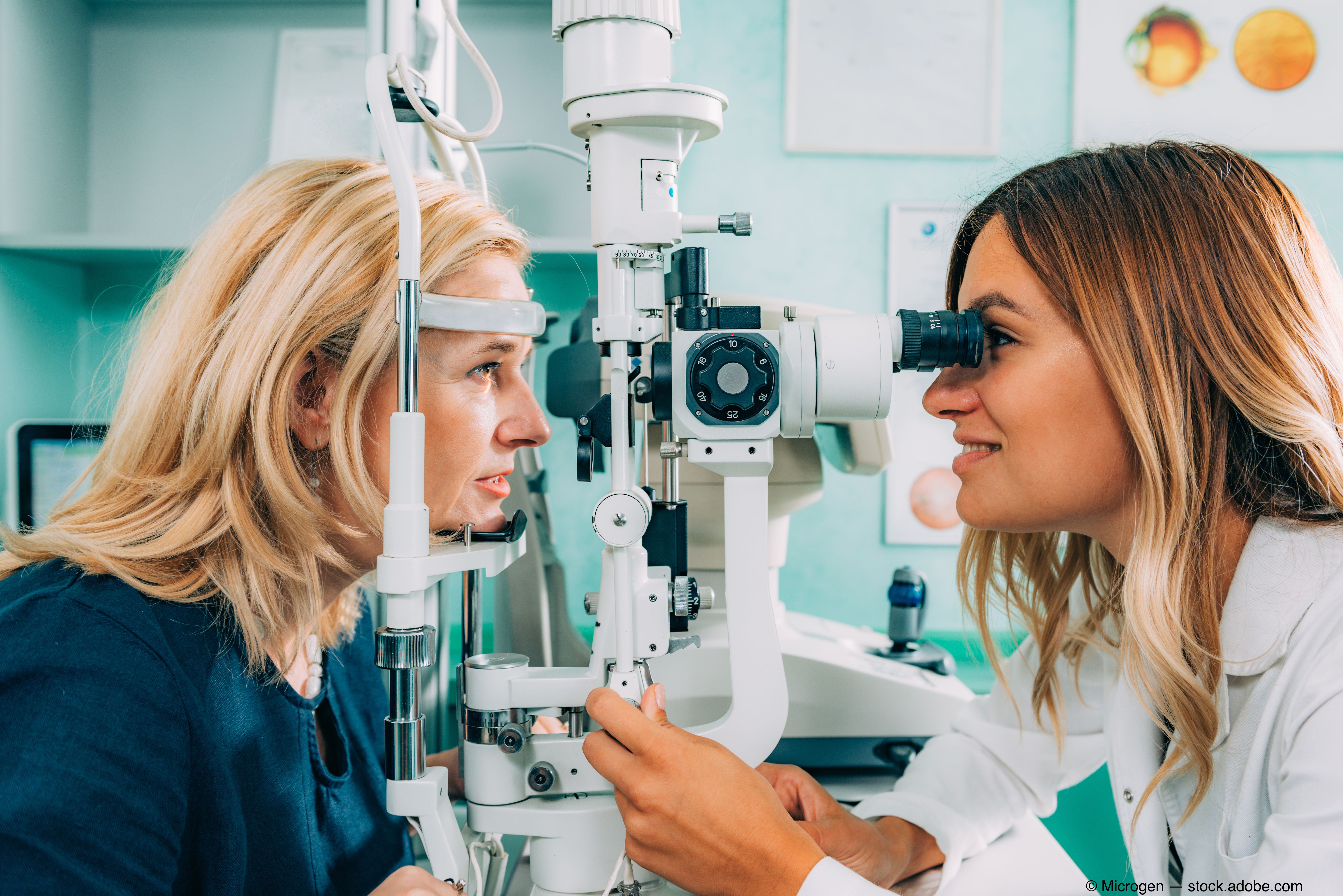
In fact, the most important tool with which I treat or diagnose dry eye with is a slit lamp. And yet it appears that the greatest obstacle ODs have when it comes to early diagnosis and treatment of this multifactorial disease is the patient’s understanding of what is actually happening. So, although ODs can put into words what they are seeing on their lids, the patients need to see it, too.
Therefore, the previous answer of the most important tool for an OD to treat dry eye with should be amended to include a picture. A slit-lamp camera is a great compliment-however, it is not always needed. Rather, ODs can download photos of similar patients’ conditions and show their patients what they are seeing. Or snap a picture through the slit lamp’s ocular with a smartphone.
Demonstrate normal function to patients and compare that to their abnormality. This can now be seen in real-time at the lids. Visualizing the lids and monitoring the progression of any meibomium gland changes is not a bourgeois diagnostic tool.
Related: Incorporating meibomian gland imaging
An OD’s job
I would argue that with that much like topography, meibography is an essential tool to help visualize changes in patients’ lid morphology. There are devices to fit an OD’s practice style and treatment that can be brought in for ease of use. Some may question if just showing the changes-without symptoms-will move patients to want to treat their lids.
Well, Doctor, that is an OD’s job. ODs must ensure patients understand the ramifications and long-term consequences, and not give them an option that is not prophylactic.
"You see it, treat it.”
ODs cannot provide patients opportunities to enhance their lives with new refractive technology if they do not first “see” what pitfalls lie ahead.
Read more comanagement articles here
References:
1. Korb DR, Blackie CA. “Dry Eye” Is the Wrong Diagnosis for Millions. Optom Vis Sci. 2015 Sep;92(9):e350-4.

Newsletter
Want more insights like this? Subscribe to Optometry Times and get clinical pearls and practice tips delivered straight to your inbox.