Cotton wool spot appears in patient with COVID-19
This case exhibits retinal correlations with the novel coronavirus


The COVID-19 pandemic continues to be arguably one of the most significant occurrences in the history of humanity. Although hope is on the horizon with vaccines, a degree of uncertainty remains regarding who will have adverse effects from the virus.
The elderly seem to be faring far worse than younger individuals. However, I know a gentleman over 80 years who contracted COVID-19 and was unaware the entire time he had it. I know what you’re thinking: he just hit a false positive. However, he went back and was tested for the antibody just for assurance, and it was confirmed he has COVID-19 antibodies.
On the other hand, a friend who is in his early 40s contracted the virus and was hospitalized with blood clots in his lungs. He stayed for several days and received serial anti-stroke medication injections. A day after being discharged, he returned to the hospital because his lung function was such that he couldn’t speak without rest. Thankfully, he is doing much better now.
Although determining factors have been identified, no one knows with 100% certainty who will fare better or worse. As for the list of possible COVID-19 complications, the eyes are not an exception. I recall when the media had a small heyday with “pink eye” as a sign of the disease. Of course it makes complete sense that COVID- 19 could make its way to the conjunctiva. But I’m not talking about conjunctivitis. I’m talking about the retina.
The retina makes sense as a potential site for COVID-19-related complications. If a virus can cause blood clots and subsequent ischemia in the lungs, then, in principle, these effects can happen anywhere there are blood vessels, including the retina.
Case example
This clinical fact was recently evidenced to me by a conversation with a colleague and friend. We will call her G. G is 51 years old and recently recovered from COVID- 19. Her pertinent negatives include the fact that she is not a diabetic and does not have hypertension, high cholesterol, or any other blood disorder. This is a reliable history because she keeps up with routine doctor visits and blood work. When she contracted the virus, she felt weak and winded. She also eventually lost her sense of taste and smell, as was common with the virus. She never had to be admitted to the hospital, and, after a couple of weeks, she was back on her feet.
Several weeks later, G started playing catch-up. This included visiting her optometrist for a comprehensive eye examination. She is a low hyperope and wears multifocal soft contact lenses. She went in with no complaints but left with a bit of a surprise.
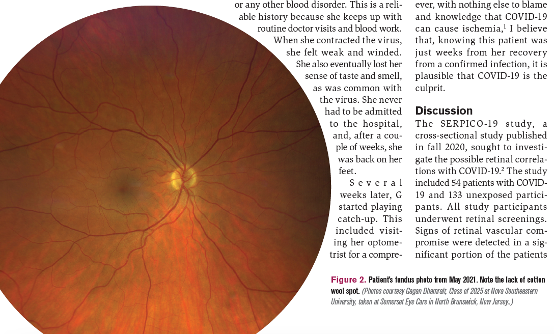
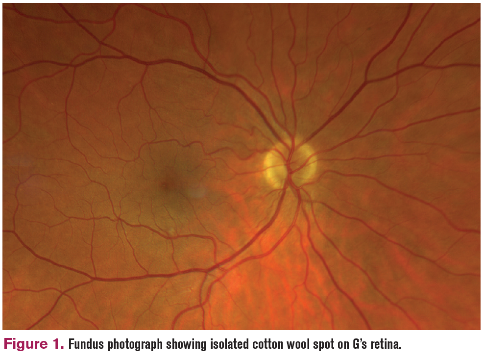
Her retinal examination showed an isolated cotton wool spot near her inferior arcade vessels in her right eye (Figure 1). It was quite small and frankly a good pick-up. It is unlikely that this particular finding will have ocular implications for her. It will likely resolve on its own. However, with nothing else to blame and knowledge that COVID-19 can cause ischemia,1 I believe that, knowing this patient was just weeks from her recovery from a confirmed infection, it is plausible that COVID-19 is the culprit.

Discussion
The SERPICO-19 study, a cross-sectional study published in fall 2020, sought to investigate the possible retinal correlations with COVID-19.2 The study included 54 patients with COVID- 19 and 133 unexposed participants. All study participants underwent retinal screenings. Signs of retinal vascular compromise were detected in a significant portion of the patients with COVID-19 wing of the study. Hemorrhages occurred in 9.25% of this population. Cotton wool spots occurred in 7.4%. Dilated retinal veins occurred in 27.7%, and vascular tortuosity occurred in 12.9%. Realizing that, for obvious reasons, the sample size was small, these percentages are still significant and support the need for further research. In fact, dilated retinal veins were associated with the degree of disease severity. This could have a possible implication for staging and treating patients with COVID-19.
My friend and colleague G inspired me to read up on COVID- 19 and the human eye. Studies such as SERPICO-19 may have implications for future treatment of patients with COVID-19. How much more of a future? Hopefully not that much.
References
1. Mahammedi A, Saba L, Vagal A, et al. Imaging in neurologic disease in hospitalized patients with COVID-19: an Italian multicenter retrospective observational study. Radiology. 2020;297(2):e270-e273. doi:10.1148/ radiol.2020201933
2. Invernizzi A, Torre A, Parrulli S, et al. Retinal findings in patients with COVID-19: results from the SERPICO-19 study. EClinicalMedicine. 2020;27:100550. doi:10.1016/j. eclinm.2020.100550

Newsletter
Want more insights like this? Subscribe to Optometry Times and get clinical pearls and practice tips delivered straight to your inbox.