What’s new in low vision rehabilitation
Offer patients independence and improve quality of life by incorporating low vision services
The promising effects of low vision rehabilitation (LVR) have been around since the 13th century when Marco Polo discovered the elderly in China using magnifiers to help them read.1 From there, ideas, theories, and research have developed into techniques and devices eye care practitioners use today in LVR.
At times, it is frustrating for practitioners and patients, when developments haven’t moved fast enough; however, recent research is giving hope to practitioners to be able to practice better, give patients more promising tools, and continue to push LVR research.
The biggest barriers to most of these advancements are cost and accessibility. Other barriers are the lack of referrals from optometrists and ophthalmologists and the resistance of eyecare practitioners to practice even basic low vision in their offices.2 However, the hope is that the knowledge and expansion of low vision research will change the accessibility of these devices and resources to low vision patients and eye care providers.
Magnifiers
Today, technology pushes LVR to new limits with advancements in everyday low vision devices allowing more affordable options for patients. LED lighting, for example, has offered patients a brighter and cleaner light source with their devices, lasting longer than the previously used halogen lighting. With LED lighting, the bulb lasts virtually forever, and a simple battery replacement makes the lighting as good as new. Illuminated magnifiers have therefore become more affordable and more sustainable.
Moving from halogen light sources with a bulb to LED lighting involves a bulb that does not need to be changed and allows a better light source for patients to improve contrast sensitivity. With this technology, manufacturers can offer limited lifetime warranties for these products. Colored filters can be added to these magnifiers’ light sources when white, bright light isn’t ideal for the patient.
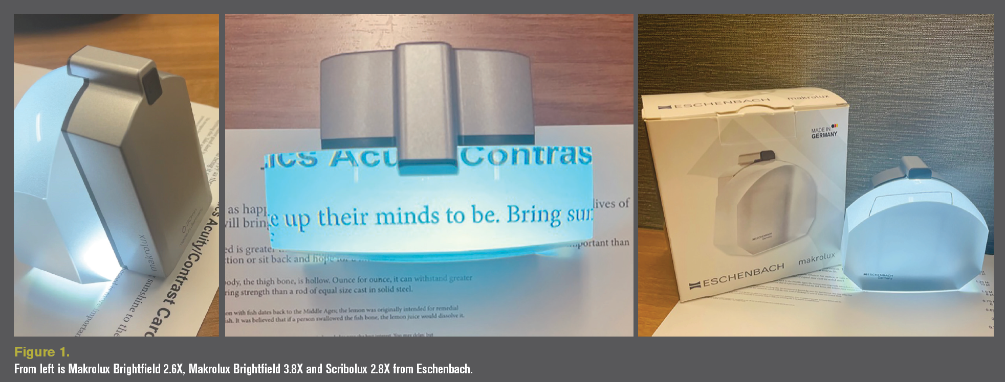
The versatility of dome magnifiers has always been popular with patients and providers, but the new illuminated options have made this favorite, simple device even better. For example, Makrolux Brightfield (Figure 1) and Scribolux magnifiers by Eschenbach offer more with optics, lighting, and functionality. These devices offer better illumination with less distortion and a larger viewing area. Although still low in magnification, these devices allow patients to read small to large print with more ease, especially because of the tilted angle for better ergonomics. Scribolux is considered a stand magnifier, but the ability to write under this magnifier makes life a bit easier, especially for students with homework. Another great feature of these illuminated magnifiers is the ability to power themselves down after 30 minutes of nonuse.
Head-mounted devices
With the events of the COVID-19 pandemic, these charts will offer low vision practitioners better options for telehealth. For example, IrisVision Live is the only smart visual assistive device that uses virtual reality (VR) to help patients with low vision to see. It also offers a portal for patients and doctors to monitor their own vision at home, incorporating visual acuity testing, visual field testing, and more. This could allow low vision practitioners not only to monitor vision for patients who don’t have the ability to come into the office on a regular basis but also to practice social distancing in everyday optometry practices.
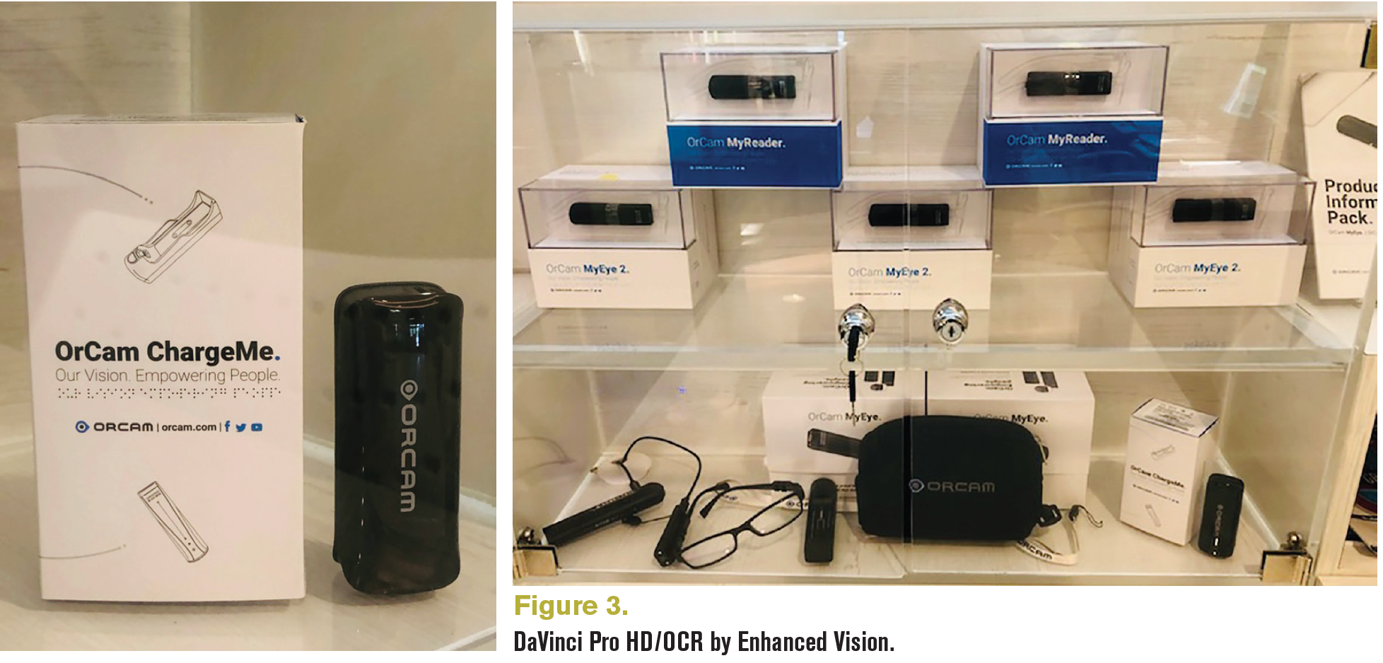
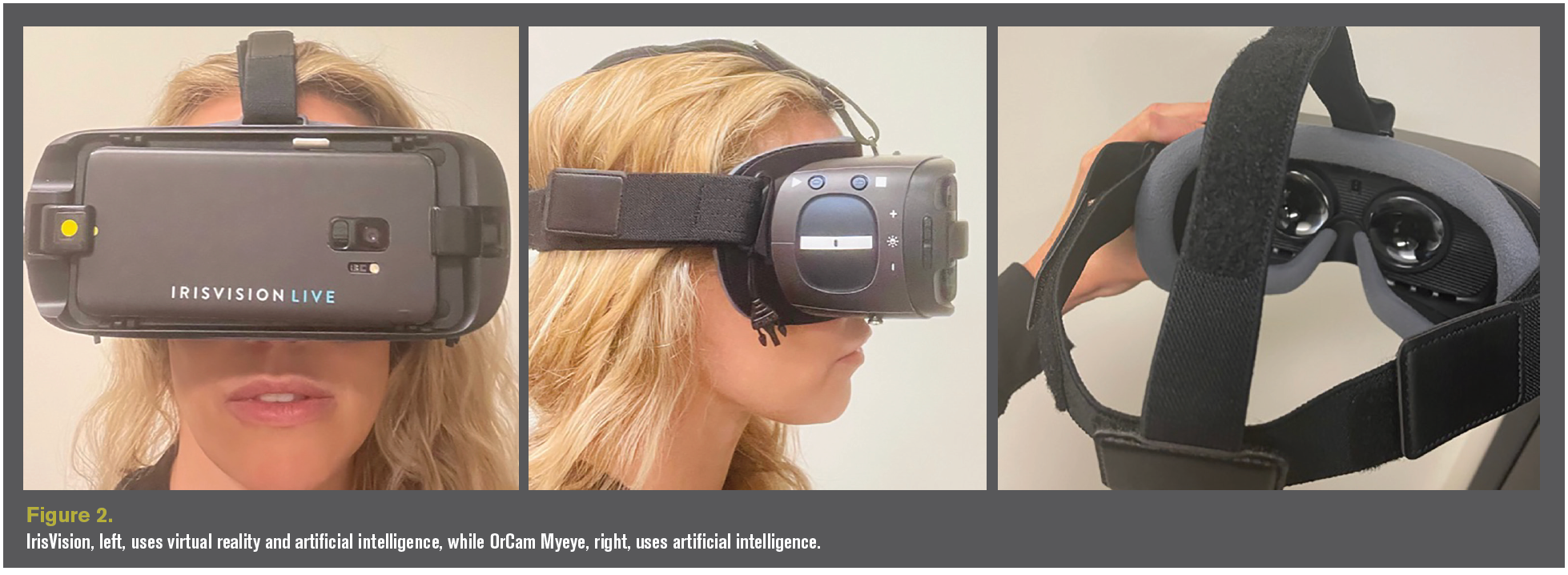
Head-mounted devices such as IrisVision (Figure 2) and OrCam products (Figure 3) use VR and/or artificial intelligence (AI) by serving as a total device set or as an attachment to the patient’s glasses. VR or augmented reality helps patients who are blind and visually impaired by using a camera that displays an image on a large screen. This allows for magnification, and can alter the image to make it easier for the viewer to see, controlling the illumination, contrast, or magnification with the push of a button or the sound of the user’s voice.
AI allows the patient to ask the device or point to something in the patient’s visual field, and the technology will tell the patient what they are looking at, such as a newspaper article they want to have the device read to them or what door to enter when they are walking down a hallway.
CCTVs
Closed-circuit TVs (CCTVs) have also come a long way. Not only are they slimmer and more attractive, and have better clarity with high-definition (HD) technology, more portable options are available as are options with optical character recognition or optical character recognition OCR technology. For students with low vision, these novel changes make it easier for them to move from place to place without lugging a ton of devices and are more socially acceptable. Such advancements make these devices more suitable for different work environments, allowing individuals to work more independently without many changes to their workspaces. They allow individuals with low vision to streamline their work needs away from the office.
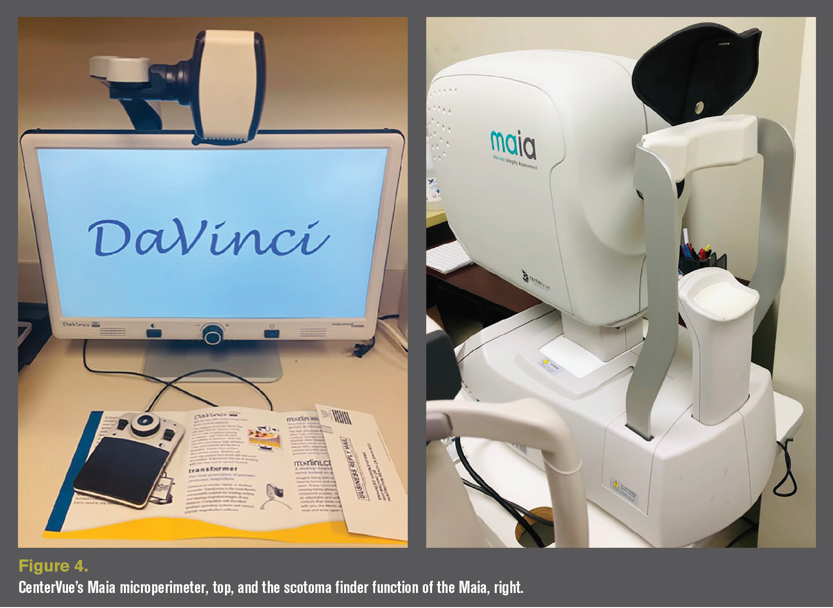
For example, Enhanced Vision’s Acrobat HD series, and Transformer HD with Wi-Fi allow for portable systems that are lightweight and provide high-definition clarity in magnification at distance and near. These systems are good choices for students in a classroom setting. DaVinci HD/OCR (Figure 4) and Pro devices offer a great combination of a CCTV that offers magnification at both distance and near, optical quality OCR technology, and a slim, sleek design that works well in an everyday office setting at work or at home.
Portable devices have advanced and have been pushed to improve because of growing competition from portable tablets, laptops, and smartphones. These devices offer slimmer, lightweight options with more magnification, better contrast, and larger viewing areas than smartphones and tablets.
Smart technology
Smart technology has been one of the most highlighted topics for low vision advancement over the last 10 years. The smartphone and its everyday applications make it more accessible to help with travel, grocery shopping, reading, or shopping online. Smartphone apps can run other devices in the home, such as lights, ovens, TVs, and other utilities. New smart devices such as Google Home and Amazon Echo allow individuals to just tell “Alexa” a command, and the task is complete.
YouTube resources
YouTube and blogs are where eye care practitioners can keep themselves updated on the latest technology and resources for patients with low vision and who are blind. Not only do these resources provide information on the most useful and new low vision devices, apps, smart technology, and helpful hints to navigate the sighted world, but they also offer inspiration.
Influencers have also been able to make a career helping others and helping themselves. These individuals offer a view into their world and how and what they struggle with on an everyday basis. Two favorites are The Blind Life by Sam Seely and Molly Burke’s The Blind Girl. Both influencers are visually impaired and discuss all topics relating to helping visually impaired people. In addition, they offer information to sighted people on what they can do and say to help the visually impaired to make them feel part of society without discrimination.
Microperimetry
Microperimetry is used by researchers and low vision practitioners to better understand the impact they can have on managing and treating macular conditions. A microperimeter uses scanning laser ophthalmoscopy to capture the patient’s responses to light stimuli at certain retinal locations and map them on a fundus photo to identify damaged and healthy retinal tissue.3
Microperimeters are used in several ways by eye care professionals. One is to test macular integrity, function, and sensitivity when assessing macular disease such as age-related macular degeneration (AMD). By performing these functions, microperimeters can help treat macular disease, predict earlier disease progression, and evaluate macular integrity and sensitivity after macular surgery. Next, the instrument can be used to help find a preferred retinal locus to improve eccentric viewing training. A microperimeter also offers biofeedback training to help patients stabilize their preferred retinal locus to help improve their efficiency with eccentric viewing. CenterVue’s Maia (Figure 5) is the preferred microperimeter because of its wide range of retinal sensitivity and biofeedback training function.3
Stem cell and genetic therapies
The advances in technology mentioned above help patients with day-to-day to activities and have changed the lives of individuals who are visually impaired or blind. However, biotechnology research has brought hope to many patients for a future where they are not as dependent on these devices and could possibly have their vision restored.
Stem cell therapy and gene therapy differ. To understand stem cell therapy, it is important to understand what stem cells are. Mayo Clinic describes stem cells as “the body’s raw materials—cells from which all other cells with specialized functions are generated.”8 Stem cells generate naturally in the body and in doing so, create “daughter cells that become new stem cells (self-renewal) or become specialized cells (differentiation) with a more specific function—no other cell in the body has the natural ability to generate new cell types.”4
Different types of stem cells exist: embryonic blood cells, adult stem cells, and perinatal blood cells. Embryonic blood cells are cells derived from 3- to 5-day-old embryos. These are blastocytes which contain about 150 cells. A blastocyte or pluripotent cell has the ability to change into any type of cell in the body, which gives it the most versatility of all stem cells.4 But harvesting embryonic blood cells is controversial. Another option is perinatal blood cells, which come from amniotic fluid and umbilical cord blood.
Adult stem cells are available but have less versatility and often can come from 1 source, typically bone marrow. However, scientists have been working on using gene therapy to change adult stem cells to work more like embryonic stem cells.4 This therapy has a lot of potential to help patients with dysfunctional retinal tissue such as AMD, Stargardt maculopathy, or retinitis pigmentosa.
Naturally occurring stem cells found in mice occur at the ciliary margin as well.5 Anterior segment disorders have made the biggest noise recently because of naturally occurring limbal stem cells. Cells located adjacent to Schwalbe’s line may also contain stem cells that could regenerate endothelial cells, which could decrease the need for corneal transplants in patients with Fuchs endothelial dystrophy.9 Research using embryonic stem cells to regenerate trabecular meshwork may help patients with glaucoma.5
Mayo Clinic describes stem cell therapy as regenerative medicine that “promotes the repair response of diseased, dysfunctional, or injured tissue using stem cells or their derivatives.”8 Scientists will harvest stem cells from one of the above sources and grow them in a lab where they are manipulated into specialized cells that can be injected into diseased tissue to help regenerate new healthy tissue. Although it seems this therapy should be the resolution to fixing diseased tissue, there are drawbacks, including the lack of control associated with stem cell growth once it is injected, and the patient’s immune system attacking the new cells.4
Gene therapy offers another solution to help patients who are visually impaired, specifically patients with inherited retinal diseases. The National Institutes of Health’s US National Library of Medicine describes gene therapy as an experimental therapy that introduces gene material into diseased cells to compensate for abnormal genes or change or promote the protein makeup of the cells.10 Injected genes are carried by a vector, usually a modified virus, to infect the cells that need to be genetically changed.6
Two entities have been the driving forces in genetic testing and gene therapy in treatment of ocular disease. In partnership with Blueprint Genetics and InformedDNA, the Foundation Fighting Blindness through its platform My Retina Tracker Program as well as Spark Therapeutics’ ID Your IRD program have made genetic testing easily accessible to practitioners as well as patients. Both platforms offer free genetic testing and free genetic counseling to patients with inherited retinal diseases or suspected inherited retinal diseases.
Once genetic testing is completed and genetic counseling is offered, the companies will inform individuals of available clinical trials. If the patient chooses to be a part of these trials, the companies can connect them with the researchers. It is as easy as directing your patients to these websites or registering your patients in your office: https:// www.fightingblindness.org/my-retina-tracker-registry and https://www.invitae.com/en/idyourird/
Conclusion
In conclusion, LVR has improved in the past 10 years and has had a significant impact on the lives of patients who are blind or visually impaired. These advancements should encourage eye care professionals to seek out how to incorporate these helpful aids and resources into their everyday practices or to refer these patients to another practitioner to provide them with the best opportunities to live their best lives.
References
1. Legge GE. Reading digital with low vision. Visible Lang. 2016;50(2):102-125.
2. Malkin AG, Ross NC, Chat TL, Protosow K, Bittner AK. US optometrists’ reported practices and perceived barriers for low vision care for mild visual loss. Optom Vis Sci. 2020;97(1):45- 51. doi:10.1097/OPX.0000000000001468
3. Stuart A. Expanded role for microperimetry in visual rehabilitation. EyeNet Magazine. April 2013. Accessed July 2, 2021. https://www.aao.org/eyenet/article/expanded-role-microperimetry-in-visual-rehabilitat
4. Mayo Clinic staff. Stem cells: what they are and what they do. Mayo Clinic. June 28, 2019. Accessed July 2, 2021. https://www.mayoclinic.org/tests-procedures/bone-marrow-transplant/in-depth/stem-cells/art-20048117
5. Levin LA, Ritch R, Richards JE, Borrás T. Stem cell therapy for ocular disorders. Arch Ophthalmol. 2004;122(4):621-27. doi:10.1001/archopht.122.4.621
6. What is gene therapy? MedlinePlus. Updated May 12, 2021. Accessed July 28, 2021. https://medlineplus.gov/genetics/ understanding/therapy/genetherapy/
Newsletter
Want more insights like this? Subscribe to Optometry Times and get clinical pearls and practice tips delivered straight to your inbox.